[English] 日本語
 Yorodumi
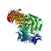
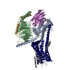
- PDB-7jvp: Cryo-EM structure of SKF-83959-bound dopamine receptor 1 in compl... -
+ Open data
- Basic information
| Entry | Database: PDB / ID: 7jvp | |||||||||||||||
|---|---|---|---|---|---|---|---|---|---|---|---|---|---|---|---|---|
| Title | Cryo-EM structure of SKF-83959-bound dopamine receptor 1 in complex with Gs protein | |||||||||||||||
 Components Components |
| |||||||||||||||
 Keywords Keywords | SIGNALING PROTEIN / Dopamine receptor 1 / Gi protein / SKF-83959 | |||||||||||||||
| Function / homology |  Function and homology information Function and homology informationdopamine neurotransmitter receptor activity, coupled via Gs / dopamine neurotransmitter receptor activity / cerebral cortex GABAergic interneuron migration / operant conditioning / Dopamine receptors / regulation of dopamine uptake involved in synaptic transmission / modification of postsynaptic structure / dopamine binding / heterotrimeric G-protein binding / G protein-coupled receptor complex ...dopamine neurotransmitter receptor activity, coupled via Gs / dopamine neurotransmitter receptor activity / cerebral cortex GABAergic interneuron migration / operant conditioning / Dopamine receptors / regulation of dopamine uptake involved in synaptic transmission / modification of postsynaptic structure / dopamine binding / heterotrimeric G-protein binding / G protein-coupled receptor complex / peristalsis / regulation of dopamine metabolic process / sensitization / grooming behavior / phospholipase C-activating dopamine receptor signaling pathway / positive regulation of neuron migration / habituation / dopamine transport / positive regulation of potassium ion transport / astrocyte development / conditioned taste aversion / striatum development / dentate gyrus development / maternal behavior / arrestin family protein binding / non-motile cilium / long-term synaptic depression / mating behavior / adult walking behavior / ciliary membrane / G protein-coupled dopamine receptor signaling pathway / temperature homeostasis / D-glucose import / transmission of nerve impulse / dopamine metabolic process / PKA activation in glucagon signalling / behavioral response to cocaine / hair follicle placode formation / G protein-coupled receptor signaling pathway, coupled to cyclic nucleotide second messenger / developmental growth / neuronal action potential / G-protein alpha-subunit binding / D1 dopamine receptor binding / intracellular transport / behavioral fear response / renal water homeostasis / Hedgehog 'off' state / prepulse inhibition / adenylate cyclase-activating adrenergic receptor signaling pathway / activation of adenylate cyclase activity / GABA-ergic synapse / cellular response to glucagon stimulus / synapse assembly / presynaptic modulation of chemical synaptic transmission / adenylate cyclase activator activity / regulation of insulin secretion / response to amphetamine / positive regulation of synaptic transmission, glutamatergic / positive regulation of release of sequestered calcium ion into cytosol / trans-Golgi network membrane / synaptic transmission, glutamatergic / G protein-coupled receptor activity / long-term synaptic potentiation / negative regulation of inflammatory response to antigenic stimulus / regulation of protein phosphorylation / bone development / visual learning / G-protein beta/gamma-subunit complex binding / adenylate cyclase-activating G protein-coupled receptor signaling pathway / Olfactory Signaling Pathway / Activation of the phototransduction cascade / G protein activity / G beta:gamma signalling through PLC beta / Presynaptic function of Kainate receptors / Thromboxane signalling through TP receptor / cilium / G protein-coupled acetylcholine receptor signaling pathway / G-protein activation / platelet aggregation / Activation of G protein gated Potassium channels / Inhibition of voltage gated Ca2+ channels via Gbeta/gamma subunits / memory / Prostacyclin signalling through prostacyclin receptor / Glucagon signaling in metabolic regulation / G beta:gamma signalling through CDC42 / cognition / G beta:gamma signalling through BTK / ADP signalling through P2Y purinoceptor 12 / Sensory perception of sweet, bitter, and umami (glutamate) taste / Synthesis, secretion, and inactivation of Glucagon-like Peptide-1 (GLP-1) / photoreceptor disc membrane / Glucagon-type ligand receptors / Adrenaline,noradrenaline inhibits insulin secretion / Vasopressin regulates renal water homeostasis via Aquaporins / G alpha (z) signalling events / vasodilation / protein import into nucleus / Glucagon-like Peptide-1 (GLP1) regulates insulin secretion / cellular response to catecholamine stimulus / ADORA2B mediated anti-inflammatory cytokines production Similarity search - Function | |||||||||||||||
| Biological species |  Homo sapiens (human) Homo sapiens (human)synthetic construct (others) | |||||||||||||||
| Method | ELECTRON MICROSCOPY / single particle reconstruction / cryo EM / Resolution: 2.9 Å | |||||||||||||||
 Authors Authors | Zhuang, Y. / Xu, P. / Mao, C. / Wang, L. / Krumm, B. / Zhou, X.E. / Huang, S. / Liu, H. / Cheng, X. / Huang, X.-P. ...Zhuang, Y. / Xu, P. / Mao, C. / Wang, L. / Krumm, B. / Zhou, X.E. / Huang, S. / Liu, H. / Cheng, X. / Huang, X.-P. / Sheng, D.-D. / Xu, T. / Liu, Y.-F. / Wang, Y. / Guo, J. / Jiang, Y. / Jiang, H. / Melcher, K. / Roth, B.L. / Zhang, Y. / Zhang, C. / Xu, H.E. | |||||||||||||||
| Funding support |  China, China,  United States, 4items United States, 4items
| |||||||||||||||
 Citation Citation |  Journal: Cell / Year: 2021 Journal: Cell / Year: 2021Title: Structural insights into the human D1 and D2 dopamine receptor signaling complexes. Authors: Youwen Zhuang / Peiyu Xu / Chunyou Mao / Lei Wang / Brian Krumm / X Edward Zhou / Sijie Huang / Heng Liu / Xi Cheng / Xi-Ping Huang / Dan-Dan Shen / Tinghai Xu / Yong-Feng Liu / Yue Wang / ...Authors: Youwen Zhuang / Peiyu Xu / Chunyou Mao / Lei Wang / Brian Krumm / X Edward Zhou / Sijie Huang / Heng Liu / Xi Cheng / Xi-Ping Huang / Dan-Dan Shen / Tinghai Xu / Yong-Feng Liu / Yue Wang / Jia Guo / Yi Jiang / Hualiang Jiang / Karsten Melcher / Bryan L Roth / Yan Zhang / Cheng Zhang / H Eric Xu /   Abstract: The D1- and D2-dopamine receptors (D1R and D2R), which signal through G and G, respectively, represent the principal stimulatory and inhibitory dopamine receptors in the central nervous system. D1R ...The D1- and D2-dopamine receptors (D1R and D2R), which signal through G and G, respectively, represent the principal stimulatory and inhibitory dopamine receptors in the central nervous system. D1R and D2R also represent the main therapeutic targets for Parkinson's disease, schizophrenia, and many other neuropsychiatric disorders, and insight into their signaling is essential for understanding both therapeutic and side effects of dopaminergic drugs. Here, we report four cryoelectron microscopy (cryo-EM) structures of D1R-G and D2R-G signaling complexes with selective and non-selective dopamine agonists, including two currently used anti-Parkinson's disease drugs, apomorphine and bromocriptine. These structures, together with mutagenesis studies, reveal the conserved binding mode of dopamine agonists, the unique pocket topology underlying ligand selectivity, the conformational changes in receptor activation, and potential structural determinants for G protein-coupling selectivity. These results provide both a molecular understanding of dopamine signaling and multiple structural templates for drug design targeting the dopaminergic system. | |||||||||||||||
| History |
|
- Structure visualization
| Movie |
 Movie viewer Movie viewer |
|---|---|
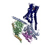
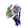
| Structure viewer | Molecule:  Molmil Molmil Jmol/JSmol Jmol/JSmol |
- Downloads & links
- Download
| PDBx/mmCIF format |  7jvp.cif.gz 7jvp.cif.gz | 212.6 KB | Display |  PDBx/mmCIF format PDBx/mmCIF format |
|---|---|---|---|---|
| PDB format |  pdb7jvp.ent.gz pdb7jvp.ent.gz | 160.7 KB | Display |  PDB format PDB format |
| PDBx/mmJSON format |  7jvp.json.gz 7jvp.json.gz | Tree view |  PDBx/mmJSON format PDBx/mmJSON format | |
| Others |  Other downloads Other downloads |
-Validation report
| Summary document |  7jvp_validation.pdf.gz 7jvp_validation.pdf.gz | 1.3 MB | Display |  wwPDB validaton report wwPDB validaton report |
|---|---|---|---|---|
| Full document |  7jvp_full_validation.pdf.gz 7jvp_full_validation.pdf.gz | 1.3 MB | Display | |
| Data in XML |  7jvp_validation.xml.gz 7jvp_validation.xml.gz | 33.8 KB | Display | |
| Data in CIF |  7jvp_validation.cif.gz 7jvp_validation.cif.gz | 51.1 KB | Display | |
| Arichive directory |  https://data.pdbj.org/pub/pdb/validation_reports/jv/7jvp https://data.pdbj.org/pub/pdb/validation_reports/jv/7jvp ftp://data.pdbj.org/pub/pdb/validation_reports/jv/7jvp ftp://data.pdbj.org/pub/pdb/validation_reports/jv/7jvp | HTTPS FTP |
-Related structure data
| Related structure data |  22509MC  7jv5C  7jvqC  7jvrC M: map data used to model this data C: citing same article ( |
|---|---|
| Similar structure data |
- Links
Links
- Assembly
| Deposited unit | 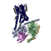
|
|---|---|
| 1 |
|
- Components
-Guanine nucleotide-binding protein ... , 3 types, 3 molecules ABG
| #2: Protein | Mass: 45624.352 Da / Num. of mol.: 1 Source method: isolated from a genetically manipulated source Source: (gene. exp.)  Homo sapiens (human) / Gene: GNAS, GNAS1, GSP / Production host: Homo sapiens (human) / Gene: GNAS, GNAS1, GSP / Production host:  |
|---|---|
| #3: Protein | Mass: 39077.715 Da / Num. of mol.: 1 Source method: isolated from a genetically manipulated source Source: (gene. exp.)  Homo sapiens (human) / Gene: GNB1 / Production host: Homo sapiens (human) / Gene: GNB1 / Production host:  |
| #4: Protein | Mass: 7432.554 Da / Num. of mol.: 1 Source method: isolated from a genetically manipulated source Source: (gene. exp.)  Homo sapiens (human) / Gene: GNG2 / Production host: Homo sapiens (human) / Gene: GNG2 / Production host:  |
-Protein / Antibody , 2 types, 2 molecules RN
| #1: Protein | Mass: 55668.707 Da / Num. of mol.: 1 Source method: isolated from a genetically manipulated source Source: (gene. exp.)  Homo sapiens (human) / Gene: DRD1 / Production host: Homo sapiens (human) / Gene: DRD1 / Production host:  |
|---|---|
| #5: Antibody | Mass: 14845.516 Da / Num. of mol.: 1 Source method: isolated from a genetically manipulated source Source: (gene. exp.) synthetic construct (others) / Production host:  |
-Non-polymers , 3 types, 12 molecules 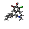


| #6: Chemical | ChemComp-SK9 / ( | ||
|---|---|---|---|
| #7: Chemical | ChemComp-CLR / #8: Chemical | ChemComp-PLM / |
-Details
| Has ligand of interest | Y |
|---|---|
| Has protein modification | Y |
-Experimental details
-Experiment
| Experiment | Method: ELECTRON MICROSCOPY |
|---|---|
| EM experiment | Aggregation state: PARTICLE / 3D reconstruction method: single particle reconstruction |
- Sample preparation
| Component | Name: SKF-81297-bound dopamine receptor 1 in complex with Gi protein Type: COMPLEX / Entity ID: #1-#5 / Source: RECOMBINANT |
|---|---|
| Source (natural) | Organism:  Homo sapiens (human) Homo sapiens (human) |
| Source (recombinant) | Organism:  |
| Buffer solution | pH: 7.2 |
| Specimen | Embedding applied: NO / Shadowing applied: NO / Staining applied: NO / Vitrification applied: YES |
| Vitrification | Cryogen name: ETHANE |
- Electron microscopy imaging
| Experimental equipment |  Model: Titan Krios / Image courtesy: FEI Company |
|---|---|
| Microscopy | Model: FEI TITAN KRIOS |
| Electron gun | Electron source:  FIELD EMISSION GUN / Accelerating voltage: 300 kV / Illumination mode: FLOOD BEAM FIELD EMISSION GUN / Accelerating voltage: 300 kV / Illumination mode: FLOOD BEAM |
| Electron lens | Mode: BRIGHT FIELD |
| Image recording | Electron dose: 64 e/Å2 / Film or detector model: GATAN K2 BASE (4k x 4k) |
- Processing
| Software | Name: PHENIX / Version: 1.13_2998: / Classification: refinement |
|---|---|
| CTF correction | Type: PHASE FLIPPING AND AMPLITUDE CORRECTION |
| 3D reconstruction | Resolution: 2.9 Å / Resolution method: FSC 0.143 CUT-OFF / Num. of particles: 679728 / Symmetry type: POINT |
 Movie
Movie Controller



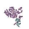

 PDBj
PDBj


