 Movie
Movie Controller


+ Open data

- Basic information
| Entry | Database: PDB / ID: 7jh7 | ||||||||||||
|---|---|---|---|---|---|---|---|---|---|---|---|---|---|
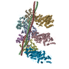
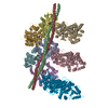
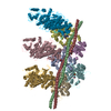
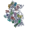
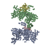
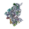
| Title | cardiac actomyosin complex | ||||||||||||
 Components Components |
| ||||||||||||
 Keywords Keywords | MOTOR PROTEIN / actomyosin | ||||||||||||
| Function / homology |  Function and homology information Function and homology informationactin-myosin filament sliding / muscle filament sliding / myosin filament / adult heart development / myosin II complex / sarcomere organization / microfilament motor activity / myosin binding / myofibril / heart contraction ...actin-myosin filament sliding / muscle filament sliding / myosin filament / adult heart development / myosin II complex / sarcomere organization / microfilament motor activity / myosin binding / myofibril / heart contraction / mesenchyme migration / cardiac muscle contraction / sarcomere / actin filament organization / filopodium / actin filament / actin filament binding / lamellipodium / cell body / calmodulin binding / positive regulation of gene expression / ATP binding / cytoplasm Similarity search - Function | ||||||||||||
| Biological species |  | ||||||||||||
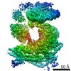
| Method | ELECTRON MICROSCOPY / helical reconstruction / cryo EM / Resolution: 3.8 Å | ||||||||||||
 Authors Authors | Galkin, V.E. / Schroeder, G.F. | ||||||||||||
| Funding support |  United States, 3items United States, 3items
| ||||||||||||
 Citation Citation | Journal: Structure / Year: 2021 Title: High-Resolution Cryo-EM Structure of the Cardiac Actomyosin Complex. Authors: Cristina Risi / Luisa U Schäfer / Betty Belknap / Ian Pepper / Howard D White / Gunnar F Schröder / Vitold E Galkin /  Abstract: Heart contraction depends on a complicated array of interactions between sarcomeric proteins required to convert chemical energy into mechanical force. Cyclic interactions between actin and myosin ...Heart contraction depends on a complicated array of interactions between sarcomeric proteins required to convert chemical energy into mechanical force. Cyclic interactions between actin and myosin molecules, controlled by troponin and tropomyosin, generate the sliding force between the actin-based thin and myosin-based thick filaments. Alterations in this sophisticated system due to missense mutations can lead to cardiovascular diseases. Numerous structural studies proposed pathological mechanisms of missense mutations at the myosin-myosin, actin-tropomyosin, and tropomyosin-troponin interfaces. However, despite the central role of actomyosin interactions a detailed structural description of the cardiac actomyosin interface remained unknown. Here, we report a cryo-EM structure of a cardiac actomyosin complex at 3.8 Å resolution. The structure reveals the molecular basis of cardiac diseases caused by missense mutations in myosin and actin proteins. | ||||||||||||
| History |
|
- Structure visualization
| Movie |
Movie viewer |
|---|---|
| Structure viewer | Molecule: MolmilJmol/JSmol |
- Downloads & links
-Download
| PDBx/mmCIF format | 7jh7.cif.gz | 801.8 KB | Display | PDBx/mmCIF format |
|---|---|---|---|---|
| PDB format | pdb7jh7.ent.gz | 626.2 KB | Display | PDB format |
| PDBx/mmJSON format | 7jh7.json.gz | Tree view | PDBx/mmJSON format | |
| Others |  Other downloads Other downloads |
-Validation report
| Summary document | 7jh7_validation.pdf.gz | 1.2 MB | Display | wwPDB validaton report |
|---|---|---|---|---|
| Full document | 7jh7_full_validation.pdf.gz | 1.2 MB | Display | |
| Data in XML | 7jh7_validation.xml.gz | 117.1 KB | Display | |
| Data in CIF | 7jh7_validation.cif.gz | 179.2 KB | Display | |
| Arichive directory | https://data.pdbj.org/pub/pdb/validation_reports/jh/7jh7ftp://data.pdbj.org/pub/pdb/validation_reports/jh/7jh7 | HTTPS FTP |
-Related structure data
| Related structure data | 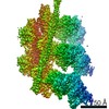 22335MC M: map data used to model this data C: citing same article ( |
|---|---|
| Similar structure data |
-Links
 PDBj
PDBj


- Assembly
| Deposited unit | 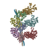
|
|---|---|
| 1 |
|
-Components
| #1: Protein | Mass: 42064.891 Da / Num. of mol.: 5 / Source method: isolated from a natural source / Source: (natural) #2: Protein | Mass: 223649.094 Da / Num. of mol.: 3 / Source method: isolated from a natural source / Source: (natural) #3: Protein | Mass: 11507.176 Da / Num. of mol.: 2 / Source method: isolated from a natural source / Source: (natural) #4: Chemical | ChemComp-ADP /   Mass: 427.201 Da / Num. of mol.: 5 / Source method: obtained synthetically / Formula: C10H15N5O10P2 / Feature type: SUBJECT OF INVESTIGATION / Comment: ADP, energy-carrying molecule*YM Mass: 427.201 Da / Num. of mol.: 5 / Source method: obtained synthetically / Formula: C10H15N5O10P2 / Feature type: SUBJECT OF INVESTIGATION / Comment: ADP, energy-carrying molecule*YM#5: Chemical | ChemComp-MG /   Mass: 24.305 Da / Num. of mol.: 5 / Source method: obtained synthetically / Formula: Mg / Feature type: SUBJECT OF INVESTIGATION Mass: 24.305 Da / Num. of mol.: 5 / Source method: obtained synthetically / Formula: Mg / Feature type: SUBJECT OF INVESTIGATIONHas ligand of interest | Y | |
|---|
-Experimental details
-Experiment
| Experiment | Method: ELECTRON MICROSCOPY |
|---|---|
| EM experiment | Aggregation state: HELICAL ARRAY / 3D reconstruction method: helical reconstruction |
- Sample preparation
| Component | Name: porcine native cardiac thin filament decorated with porcine cardiac myosin-S1 Type: COMPLEX / Entity ID: #1-#3 / Source: NATURAL |
|---|---|
| Molecular weight | Value: 492 kDa/nm / Experimental value: NO |
| Source (natural) | Organism: |
| Buffer solution | pH: 7 |
| Specimen | Conc.: 0.09 mg/ml / Embedding applied: NO / Shadowing applied: NO / Staining applied: NO / Vitrification applied: YES Details: 3 uL of 2 uM TFs were applied to glow-discharged lacey carbon grid for 1 min, gently blotted with Whatman #1 filter paper, and incubated with 2 uL of myosin-S1 [1.2 uM] for 2 min |
| Specimen support | Grid material: COPPER / Grid mesh size: 300 divisions/in. / Grid type: Quantifoil |
| Vitrification | Instrument: FEI VITROBOT MARK IV / Cryogen name: ETHANE / Humidity: 100 % / Chamber temperature: 283 K |
- Electron microscopy imaging
| Experimental equipment |  Model: Titan Krios / Image courtesy: FEI Company |
|---|---|
| Microscopy | Model: FEI TITAN KRIOS |
| Electron gun | Electron source:  FIELD EMISSION GUN / Accelerating voltage: 300 kV / Illumination mode: FLOOD BEAM FIELD EMISSION GUN / Accelerating voltage: 300 kV / Illumination mode: FLOOD BEAM |
| Electron lens | Mode: BRIGHT FIELD |
| Specimen holder | Cryogen: NITROGEN / Specimen holder model: FEI TITAN KRIOS AUTOGRID HOLDER |
| Image recording | Electron dose: 20 e/Å2 / Film or detector model: FEI FALCON III (4k x 4k) / Num. of grids imaged: 1 / Num. of real images: 2373 |
- Processing
| Software | Name: PHENIX / Version: 1.11.1_2575: / Classification: refinement | ||||||||||||||||||||||||||||||||||||
|---|---|---|---|---|---|---|---|---|---|---|---|---|---|---|---|---|---|---|---|---|---|---|---|---|---|---|---|---|---|---|---|---|---|---|---|---|---|
| EM software |
| ||||||||||||||||||||||||||||||||||||
| CTF correction | Type: PHASE FLIPPING AND AMPLITUDE CORRECTION | ||||||||||||||||||||||||||||||||||||
| Helical symmerty | Angular rotation/subunit: -166.7 ° / Axial rise/subunit: 27.3 Å / Axial symmetry: C1 | ||||||||||||||||||||||||||||||||||||
| 3D reconstruction | Resolution: 3.8 Å / Resolution method: FSC 0.143 CUT-OFF / Num. of particles: 115746 / Algorithm: BACK PROJECTION / Symmetry type: HELICAL | ||||||||||||||||||||||||||||||||||||
| Atomic model building | Protocol: FLEXIBLE FIT / Space: REAL Details: SWISS-MODEL web service used to create initial homology models of cardiac actin and myosin | ||||||||||||||||||||||||||||||||||||
| Refine LS restraints |
|
