 Movie
Movie Controller

[English] 日本語
 Yorodumi
- PDB-7f5b: LBD-TMD focused reconstruction of DNQX-bound GluK2-1xNeto2 complex -
+ Open data

- Basic information
| Entry | Database: PDB / ID: 7f5b | ||||||||||||||||||||||||||||||||||||
|---|---|---|---|---|---|---|---|---|---|---|---|---|---|---|---|---|---|---|---|---|---|---|---|---|---|---|---|---|---|---|---|---|---|---|---|---|---|
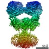
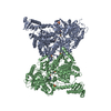
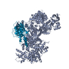
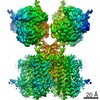
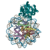
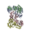
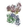
| Title | LBD-TMD focused reconstruction of DNQX-bound GluK2-1xNeto2 complex | ||||||||||||||||||||||||||||||||||||
 Components Components |
| ||||||||||||||||||||||||||||||||||||
 Keywords Keywords | MEMBRANE PROTEIN / Ionotropic glutamate receptors / Single-pass transmembrane proteins | ||||||||||||||||||||||||||||||||||||
| Function / homology |  Function and homology information Function and homology informationmossy fiber rosette / detection of cold stimulus involved in thermoception / Activation of Na-permeable kainate receptors / Activation of Ca-permeable Kainate Receptor / kainate selective glutamate receptor complex / regulation of short-term neuronal synaptic plasticity / ubiquitin conjugating enzyme binding / glutamate receptor activity / negative regulation of synaptic transmission, glutamatergic / regulation of JNK cascade ...mossy fiber rosette / detection of cold stimulus involved in thermoception / Activation of Na-permeable kainate receptors / Activation of Ca-permeable Kainate Receptor / kainate selective glutamate receptor complex / regulation of short-term neuronal synaptic plasticity / ubiquitin conjugating enzyme binding / glutamate receptor activity / negative regulation of synaptic transmission, glutamatergic / regulation of JNK cascade / neurotransmitter receptor localization to postsynaptic specialization membrane / receptor clustering / regulation of neurotransmitter receptor localization to postsynaptic specialization membrane / inhibitory postsynaptic potential / kainate selective glutamate receptor activity / extracellularly glutamate-gated ion channel activity / modulation of excitatory postsynaptic potential / ionotropic glutamate receptor complex / positive regulation of synaptic transmission / behavioral fear response / neuronal action potential / glutamate-gated receptor activity / glutamate-gated calcium ion channel activity / ionotropic glutamate receptor binding / dendrite cytoplasm / ligand-gated monoatomic ion channel activity involved in regulation of presynaptic membrane potential / presynaptic modulation of chemical synaptic transmission / hippocampal mossy fiber to CA3 synapse / SNARE binding / PDZ domain binding / transmitter-gated monoatomic ion channel activity involved in regulation of postsynaptic membrane potential / synaptic transmission, glutamatergic / regulation of membrane potential / excitatory postsynaptic potential / intracellular protein transport / regulation of long-term neuronal synaptic plasticity / postsynaptic density membrane / modulation of chemical synaptic transmission / intracellular calcium ion homeostasis / terminal bouton / positive regulation of neuron apoptotic process / neuron apoptotic process / scaffold protein binding / presynaptic membrane / chemical synaptic transmission / negative regulation of neuron apoptotic process / perikaryon / postsynaptic membrane / postsynaptic density / axon / neuronal cell body / ubiquitin protein ligase binding / synapse / dendrite / glutamatergic synapse / membrane / identical protein binding / plasma membrane Similarity search - Function | ||||||||||||||||||||||||||||||||||||
| Biological species |  | ||||||||||||||||||||||||||||||||||||
| Method | ELECTRON MICROSCOPY / single particle reconstruction / cryo EM / Resolution: 3.9 Å | ||||||||||||||||||||||||||||||||||||
 Authors Authors | He, L.L. / Gao, Y.W. / Li, B. / Zhao, Y. | ||||||||||||||||||||||||||||||||||||
| Funding support |  China, 1items China, 1items
| ||||||||||||||||||||||||||||||||||||
 Citation Citation | Journal: Nature / Year: 2021 Title: Kainate receptor modulation by NETO2. Authors: Lingli He / Jiahui Sun / Yiwei Gao / Bin Li / Yuhang Wang / Yanli Dong / Weidong An / Hang Li / Bei Yang / Yuhan Ge / Xuejun Cai Zhang / Yun Stone Shi / Yan Zhao / Abstract: Glutamate-gated kainate receptors are ubiquitous in the central nervous system of vertebrates, mediate synaptic transmission at the postsynapse and modulate transmitter release at the presynapse. In ...Glutamate-gated kainate receptors are ubiquitous in the central nervous system of vertebrates, mediate synaptic transmission at the postsynapse and modulate transmitter release at the presynapse. In the brain, the trafficking, gating kinetics and pharmacology of kainate receptors are tightly regulated by neuropilin and tolloid-like (NETO) proteins. Here we report cryo-electron microscopy structures of homotetrameric GluK2 in complex with NETO2 at inhibited and desensitized states, illustrating variable stoichiometry of GluK2-NETO2 complexes, with one or two NETO2 subunits associating with GluK2. We find that NETO2 accesses only two broad faces of kainate receptors, intermolecularly crosslinking the lower lobe of ATD, the upper lobe of LBD and the lower lobe of LBD, illustrating how NETO2 regulates receptor-gating kinetics. The transmembrane helix of NETO2 is positioned proximal to the selectivity filter and competes with the amphiphilic H1 helix after M4 for interaction with an intracellular cap domain formed by the M1-M2 linkers of the receptor, revealing how rectification is regulated by NETO2. | ||||||||||||||||||||||||||||||||||||
| History |
|
- Structure visualization
| Movie |
Movie viewer |
|---|---|
| Structure viewer | Molecule: MolmilJmol/JSmol |
- Downloads & links
-Download
| PDBx/mmCIF format | 7f5b.cif.gz | 383 KB | Display | PDBx/mmCIF format |
|---|---|---|---|---|
| PDB format | pdb7f5b.ent.gz | 289.1 KB | Display | PDB format |
| PDBx/mmJSON format | 7f5b.json.gz | Tree view | PDBx/mmJSON format | |
| Others |  Other downloads Other downloads |
-Validation report
| Arichive directory | https://data.pdbj.org/pub/pdb/validation_reports/f5/7f5bftp://data.pdbj.org/pub/pdb/validation_reports/f5/7f5b | HTTPS FTP |
|---|
-Related structure data
| Related structure data |  31464MC  7f56C  7f57C  7f59C 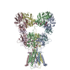 7f5aC M: map data used to model this data C: citing same article ( |
|---|---|
| Similar structure data |


-Links
 PDBj
PDBj


- Assembly
| Deposited unit | 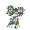
|
|---|---|
| 1 |
|
-Components
-Protein , 2 types, 5 molecules ABCDE
| #1: Protein | Mass: 102475.961 Da / Num. of mol.: 4 / Mutation: F107L Source method: isolated from a genetically manipulated source Source: (gene. exp.)  Homo sapiens (human) / References: UniProt: P42260 Homo sapiens (human) / References: UniProt: P42260#2: Protein | | Mass: 59413.652 Da / Num. of mol.: 1 Source method: isolated from a genetically manipulated source Source: (gene. exp.) Homo sapiens (human) / References: UniProt: C6K2K4 |
|---|
-Sugars , 2 types, 8 molecules 
| #3: Polysaccharide | Source method: isolated from a genetically manipulated source #4: Sugar | ChemComp-NAG /  Type: D-saccharide, beta linking / Mass: 221.208 Da / Num. of mol.: 6 / Source method: obtained synthetically / Formula: C8H15NO6 Type: D-saccharide, beta linking / Mass: 221.208 Da / Num. of mol.: 6 / Source method: obtained synthetically / Formula: C8H15NO6 |
|---|
-Non-polymers , 2 types, 2 molecules 
| #5: Chemical | ChemComp-CA /  Mass: 40.078 Da / Num. of mol.: 1 / Source method: obtained synthetically / Formula: Ca / Feature type: SUBJECT OF INVESTIGATION Mass: 40.078 Da / Num. of mol.: 1 / Source method: obtained synthetically / Formula: Ca / Feature type: SUBJECT OF INVESTIGATION |
|---|---|
| #6: Chemical | ChemComp-PGT / ( Mass: 751.023 Da / Num. of mol.: 1 / Source method: obtained synthetically / Formula: C40H79O10P / Comment: phospholipid*YM Mass: 751.023 Da / Num. of mol.: 1 / Source method: obtained synthetically / Formula: C40H79O10P / Comment: phospholipid*YM |
-Details
| Has ligand of interest | Y |
|---|---|
| Has protein modification | Y |
-Experimental details
-Experiment
| Experiment | Method: ELECTRON MICROSCOPY |
|---|---|
| EM experiment | Aggregation state: PARTICLE / 3D reconstruction method: single particle reconstruction |
- Sample preparation
| Component | Name: LBD-TMD focused reconstruction of DNQX-bound GluK2-1xNeto2 complex Type: COMPLEX / Entity ID: #1-#2 / Source: RECOMBINANT |
|---|---|
| Source (natural) | Organism: |
| Source (recombinant) | Organism: Homo sapiens (human) |
| Buffer solution | pH: 7.4 |
| Specimen | Embedding applied: NO / Shadowing applied: NO / Staining applied: NO / Vitrification applied: YES |
| Vitrification | Cryogen name: ETHANE |
- Electron microscopy imaging
| Experimental equipment |  Model: Titan Krios / Image courtesy: FEI Company |
|---|---|
| Microscopy | Model: FEI TITAN KRIOS |
| Electron gun | Electron source:  FIELD EMISSION GUN / Accelerating voltage: 300 kV / Illumination mode: FLOOD BEAM FIELD EMISSION GUN / Accelerating voltage: 300 kV / Illumination mode: FLOOD BEAM |
| Electron lens | Mode: BRIGHT FIELD |
| Image recording | Electron dose: 60 e/Å2 / Detector mode: SUPER-RESOLUTION / Film or detector model: GATAN K2 SUMMIT (4k x 4k) |
- Processing
| Software | Name: PHENIX / Version: 1.18.2_3874: / Classification: refinement | ||||||||||||||||||||||||
|---|---|---|---|---|---|---|---|---|---|---|---|---|---|---|---|---|---|---|---|---|---|---|---|---|---|
| EM software | Name: PHENIX / Category: model refinement | ||||||||||||||||||||||||
| CTF correction | Type: NONE | ||||||||||||||||||||||||
| 3D reconstruction | Resolution: 3.9 Å / Resolution method: FSC 0.143 CUT-OFF / Num. of particles: 148300 / Symmetry type: POINT | ||||||||||||||||||||||||
| Refine LS restraints |
|
