Movie
Movie Controller


+ Open data

- Basic information
| Entry | Database: PDB / ID: 7.0E+55 | ||||||||||||||||||
|---|---|---|---|---|---|---|---|---|---|---|---|---|---|---|---|---|---|---|---|
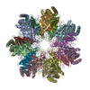
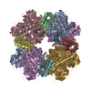
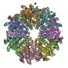
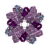
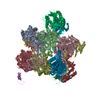
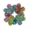
| Title | Cryo-EM structure of alpha 7 homo-tetradecamer | ||||||||||||||||||
 Components Components | Proteasome subunit alpha type-3 | ||||||||||||||||||
 Keywords Keywords | LYASE / Conformational fluctuation / double-ring / proteasome / D7 symmetry / CHAPERONE | ||||||||||||||||||
| Function / homology |  Function and homology information Function and homology informationAntigen processing: Ub, ATP-independent proteasomal degradation / Regulation of ornithine decarboxylase (ODC) / Proteasome assembly / proteasome core complex / Cross-presentation of soluble exogenous antigens (endosomes) / Somitogenesis / proteasome core complex, alpha-subunit complex / proteasome complex / Regulation of activated PAK-2p34 by proteasome mediated degradation / Autodegradation of Cdh1 by Cdh1:APC/C ...Antigen processing: Ub, ATP-independent proteasomal degradation / Regulation of ornithine decarboxylase (ODC) / Proteasome assembly / proteasome core complex / Cross-presentation of soluble exogenous antigens (endosomes) / Somitogenesis / proteasome core complex, alpha-subunit complex / proteasome complex / Regulation of activated PAK-2p34 by proteasome mediated degradation / Autodegradation of Cdh1 by Cdh1:APC/C / APC/C:Cdc20 mediated degradation of Securin / Asymmetric localization of PCP proteins / Ubiquitin-dependent degradation of Cyclin D / SCF-beta-TrCP mediated degradation of Emi1 / NIK-->noncanonical NF-kB signaling / AUF1 (hnRNP D0) binds and destabilizes mRNA / TNFR2 non-canonical NF-kB pathway / Assembly of the pre-replicative complex / Vpu mediated degradation of CD4 / Cdc20:Phospho-APC/C mediated degradation of Cyclin A / Dectin-1 mediated noncanonical NF-kB signaling / Degradation of DVL / Degradation of AXIN / Degradation of CRY and PER proteins / Hh mutants are degraded by ERAD / Activation of NF-kappaB in B cells / G2/M Checkpoints / Degradation of GLI1 by the proteasome / Hedgehog ligand biogenesis / Regulation of RUNX3 expression and activity / Autodegradation of the E3 ubiquitin ligase COP1 / Defective CFTR causes cystic fibrosis / GSK3B and BTRC:CUL1-mediated-degradation of NFE2L2 / Negative regulation of NOTCH4 signaling / APC/C:Cdh1 mediated degradation of Cdc20 and other APC/C:Cdh1 targeted proteins in late mitosis/early G1 / Hedgehog 'on' state / Vif-mediated degradation of APOBEC3G / FBXL7 down-regulates AURKA during mitotic entry and in early mitosis / Degradation of GLI2 by the proteasome / GLI3 is processed to GLI3R by the proteasome / Ubiquitin-Mediated Degradation of Phosphorylated Cdc25A / MAPK6/MAPK4 signaling / Degradation of CDH1 / Degradation of beta-catenin by the destruction complex / Oxygen-dependent proline hydroxylation of Hypoxia-inducible Factor Alpha / CDK-mediated phosphorylation and removal of Cdc6 / ABC-family protein mediated transport / CLEC7A (Dectin-1) signaling / SCF(Skp2)-mediated degradation of p27/p21 / FCERI mediated NF-kB activation / Regulation of expression of SLITs and ROBOs / Regulation of PTEN stability and activity / Interleukin-1 signaling / Orc1 removal from chromatin / Regulation of RAS by GAPs / Regulation of RUNX2 expression and activity / The role of GTSE1 in G2/M progression after G2 checkpoint / Separation of Sister Chromatids / UCH proteinases / KEAP1-NFE2L2 pathway / Downstream TCR signaling / Antigen processing: Ubiquitination & Proteasome degradation / RUNX1 regulates transcription of genes involved in differentiation of HSCs / ER-Phagosome pathway / Neddylation / proteasome-mediated ubiquitin-dependent protein catabolic process / Ub-specific processing proteases / ubiquitin protein ligase binding / extracellular exosome / nucleoplasm / nucleus / cytoplasm / cytosol Similarity search - Function | ||||||||||||||||||
| Biological species |  Homo sapiens (human) Homo sapiens (human) | ||||||||||||||||||
| Method | ELECTRON MICROSCOPY / single particle reconstruction / cryo EM / Resolution: 5.9 Å | ||||||||||||||||||
 Authors Authors | Song, C. / Murata, K. | ||||||||||||||||||
| Funding support |  Japan, 5items Japan, 5items
| ||||||||||||||||||
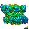
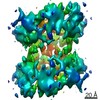
 Citation Citation | Journal: Int J Mol Sci / Year: 2021 Title: Structural Fluctuations of the Human Proteasome α7 Homo-Tetradecamer Double Ring Imply the Proteasomal α-Ring Assembly Mechanism. Authors: Chihong Song / Tadashi Satoh / Taichiro Sekiguchi / Koichi Kato / Kazuyoshi Murata / Abstract: The 20S proteasome, which is composed of layered α and β heptameric rings, is the core complex of the eukaryotic proteasome involved in proteolysis. The α7 subunit is a component of the α ring, ...The 20S proteasome, which is composed of layered α and β heptameric rings, is the core complex of the eukaryotic proteasome involved in proteolysis. The α7 subunit is a component of the α ring, and it self-assembles into a homo-tetradecamer consisting of two layers of α7 heptameric rings. However, the structure of the α7 double ring in solution has not been fully elucidated. We applied cryo-electron microscopy to delineate the structure of the α7 double ring in solution, revealing a structure different from the previously reported crystallographic model. The D7-symmetrical double ring was stacked with a 15° clockwise twist and a separation of 3 Å between the two rings. Two more conformations, dislocated and fully open, were also identified. Our observations suggest that the α7 double-ring structure fluctuates considerably in solution, allowing for the insertion of homologous α subunits, finally converting to the hetero-heptameric α rings in the 20S proteasome. | ||||||||||||||||||
| History |
|
- Structure visualization
| Movie |
Movie viewer |
|---|---|
| Structure viewer | Molecule: MolmilJmol/JSmol |
- Downloads & links
-Download
| PDBx/mmCIF format | 7e55.cif.gz | 55.2 KB | Display | PDBx/mmCIF format |
|---|---|---|---|---|
| PDB format | pdb7e55.ent.gz | 38.5 KB | Display | PDB format |
| PDBx/mmJSON format | 7e55.json.gz | Tree view | PDBx/mmJSON format | |
| Others |  Other downloads Other downloads |
-Validation report
| Arichive directory | https://data.pdbj.org/pub/pdb/validation_reports/e5/7e55ftp://data.pdbj.org/pub/pdb/validation_reports/e5/7e55 | HTTPS FTP |
|---|
-Related structure data
| Related structure data | 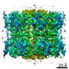 30990MC M: map data used to model this data C: citing same article ( |
|---|---|
| Similar structure data |
-Links
 PDBj
PDBj


- Assembly
| Deposited unit | 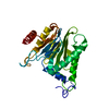
|
|---|---|
| 1 | x 14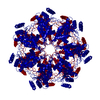
|
| 2 |
|
| 3 | 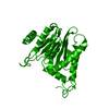
|
| Symmetry | Point symmetry: (Schoenflies symbol: D7 (2x7 fold dihedral)) |
-Components
| #1: Protein | Mass: 28469.252 Da / Num. of mol.: 1 Source method: isolated from a genetically manipulated source Source: (gene. exp.) Homo sapiens (human) / Gene: PSMA3, HC8, PSC8 / Production host:  |
|---|---|
| Has protein modification | Y |
-Experimental details
-Experiment
| Experiment | Method: ELECTRON MICROSCOPY |
|---|---|
| EM experiment | Aggregation state: PARTICLE / 3D reconstruction method: single particle reconstruction |
- Sample preparation
| Component | Name: Proteasome subunit alpha type-7 / Type: COMPLEX / Entity ID: all / Source: RECOMBINANT |
|---|---|
| Source (natural) | Organism: Homo sapiens (human) |
| Source (recombinant) | Organism: |
| Buffer solution | pH: 7.5 |
| Specimen | Conc.: 1 mg/ml / Embedding applied: YES / Shadowing applied: NO / Staining applied: NO / Vitrification applied: YES |
| Specimen support | Grid material: MOLYBDENUM / Grid mesh size: 200 divisions/in. / Grid type: Quantifoil R1.2/1.3 |
| EM embedding | Material: Ice |
| Vitrification | Cryogen name: ETHANE / Humidity: 95 % / Chamber temperature: 277 K |
- Electron microscopy imaging
| Microscopy | Model: JEOL 2200FS |
|---|---|
| Electron gun | Electron source:  FIELD EMISSION GUN / Accelerating voltage: 200 kV / Illumination mode: FLOOD BEAM FIELD EMISSION GUN / Accelerating voltage: 200 kV / Illumination mode: FLOOD BEAM |
| Electron lens | Mode: BRIGHT FIELD / Nominal magnification: 40000 X / Calibrated magnification: 45065 X / Nominal defocus max: 3000 nm / Nominal defocus min: 1000 nm / Cs: 4.2 mm / C2 aperture diameter: 40 µm / Alignment procedure: BASIC |
| Specimen holder | Cryogen: NITROGEN Specimen holder model: GATAN 626 SINGLE TILT LIQUID NITROGEN CRYO TRANSFER HOLDER Temperature (max): 77 K / Temperature (min): 76 K |
| Image recording | Average exposure time: 5 sec. / Electron dose: 40 e/Å2 / Film or detector model: DIRECT ELECTRON DE-20 (5k x 3k) |
| EM imaging optics | Energyfilter name: In-column Omega Filter / Energyfilter slit width: 20 eV |
| Image scans | Sampling size: 6.4 µm / Width: 5120 / Height: 3840 / Movie frames/image: 25 |
- Processing
| EM software |
| ||||||||||||||||||||||||||||||||
|---|---|---|---|---|---|---|---|---|---|---|---|---|---|---|---|---|---|---|---|---|---|---|---|---|---|---|---|---|---|---|---|---|---|
| CTF correction | Type: PHASE FLIPPING ONLY | ||||||||||||||||||||||||||||||||
| Symmetry | Point symmetry: D7 (2x7 fold dihedral) | ||||||||||||||||||||||||||||||||
| 3D reconstruction | Resolution: 5.9 Å / Resolution method: FSC 0.143 CUT-OFF / Num. of particles: 18388 / Algorithm: BACK PROJECTION / Symmetry type: POINT | ||||||||||||||||||||||||||||||||
| Atomic model building | B value: 531 / Protocol: FLEXIBLE FIT / Space: REAL | ||||||||||||||||||||||||||||||||
| Atomic model building | PDB-ID: 5DSV Accession code: 5DSV / Pdb chain residue range: 2-245 / Source name: PDB / Type: experimental model |