 Movie
Movie Controller


+ Open data

- Basic information
| Entry | Database: PDB / ID: 7dtt | ||||||
|---|---|---|---|---|---|---|---|
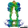
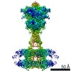
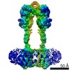
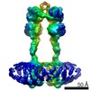
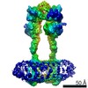
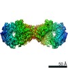
| Title | Human Calcium-Sensing Receptor bound with calcium ions | ||||||
 Components Components | Extracellular calcium-sensing receptor | ||||||
 Keywords Keywords | MEMBRANE PROTEIN / GPCR / class C / calcium sensing receptor | ||||||
| Function / homology |  Function and homology information Function and homology informationregulation of presynaptic membrane potential / bile acid secretion / chemosensory behavior / response to fibroblast growth factor / cellular response to peptide / cellular response to vitamin D / Class C/3 (Metabotropic glutamate/pheromone receptors) / positive regulation of positive chemotaxis / fat pad development / cellular response to hepatocyte growth factor stimulus ...regulation of presynaptic membrane potential / bile acid secretion / chemosensory behavior / response to fibroblast growth factor / cellular response to peptide / cellular response to vitamin D / Class C/3 (Metabotropic glutamate/pheromone receptors) / positive regulation of positive chemotaxis / fat pad development / cellular response to hepatocyte growth factor stimulus / amino acid binding / branching morphogenesis of an epithelial tube / positive regulation of calcium ion import / regulation of calcium ion transport / positive regulation of NLRP3 inflammasome complex assembly / positive regulation of vasoconstriction / anatomical structure morphogenesis / cellular response to low-density lipoprotein particle stimulus / detection of calcium ion / JNK cascade / ossification / axon terminus / response to ischemia / chloride transmembrane transport / cellular response to glucose stimulus / integrin binding / vasodilation / G protein-coupled receptor activity / intracellular calcium ion homeostasis / adenylate cyclase-inhibiting G protein-coupled receptor signaling pathway / positive regulation of insulin secretion / presynaptic membrane / cellular response to hypoxia / G alpha (i) signalling events / basolateral plasma membrane / phospholipase C-activating G protein-coupled receptor signaling pathway / G alpha (q) signalling events / transmembrane transporter binding / positive regulation of ERK1 and ERK2 cascade / apical plasma membrane / G protein-coupled receptor signaling pathway / neuronal cell body / calcium ion binding / positive regulation of cell population proliferation / positive regulation of gene expression / protein kinase binding / glutamatergic synapse / cell surface / protein homodimerization activity / identical protein binding / plasma membrane Similarity search - Function | ||||||
| Biological species |  Homo sapiens (human) Homo sapiens (human) | ||||||
| Method | ELECTRON MICROSCOPY / single particle reconstruction / cryo EM / Resolution: 3.8 Å | ||||||
 Authors Authors | Ling, S.L. / Tian, C.L. / Shi, P. / Liu, S.L. / Meng, X.Y. / Sun, D.M. / Liu, L. / Shi, C.W. | ||||||
| Funding support |  China, 1items China, 1items
| ||||||
 Citation Citation | Journal: Cell Res / Year: 2021 Title: Structural mechanism of cooperative activation of the human calcium-sensing receptor by Ca ions and L-tryptophan. Authors: Shenglong Ling / Pan Shi / Sanling Liu / Xianyu Meng / Yingxin Zhou / Wenjing Sun / Shenghai Chang / Xing Zhang / Longhua Zhang / Chaowei Shi / Demeng Sun / Lei Liu / Changlin Tian / Abstract: The human calcium-sensing receptor (CaSR) is a class C G protein-coupled receptor (GPCR) responsible for maintaining Ca homeostasis in the blood. The general consensus is that extracellular Ca is ...The human calcium-sensing receptor (CaSR) is a class C G protein-coupled receptor (GPCR) responsible for maintaining Ca homeostasis in the blood. The general consensus is that extracellular Ca is the principal agonist of CaSR. Aliphatic and aromatic L-amino acids, such as L-Phe and L-Trp, increase the sensitivity of CaSR towards Ca and are considered allosteric activators. Crystal structures of the extracellular domain (ECD) of CaSR dimer have demonstrated Ca and L-Trp binding sites and conformational changes of the ECD upon Ca/L-Trp binding. However, it remains to be understood at the structural level how Ca/L-Trp binding to the ECD leads to conformational changes in transmembrane domains (TMDs) and consequent CaSR activation. Here, we determined the structures of full-length human CaSR in the inactive state, Ca- or L-Trp-bound states, and Ca/L-Trp-bound active state using single-particle cryo-electron microscopy. Structural studies demonstrate that L-Trp binding induces the closure of the Venus flytrap (VFT) domain of CaSR, bringing the receptor into an intermediate active state. Ca binding relays the conformational changes from the VFT domains to the TMDs, consequently inducing close contact between the two TMDs of dimeric CaSR, activating the receptor. Importantly, our structural and functional studies reveal that Ca ions and L-Trp activate CaSR cooperatively. Amino acids are not able to activate CaSR alone, but can promote the receptor activation in the presence of Ca. Our data provide complementary insights into the activation of class C GPCRs and may aid in the development of novel drugs targeting CaSR. | ||||||
| History |
|
- Structure visualization
| Movie |
Movie viewer |
|---|---|
| Structure viewer | Molecule: MolmilJmol/JSmol |
- Downloads & links
-Download
| PDBx/mmCIF format | 7dtt.cif.gz | 286.9 KB | Display | PDBx/mmCIF format |
|---|---|---|---|---|
| PDB format | pdb7dtt.ent.gz | 213.6 KB | Display | PDB format |
| PDBx/mmJSON format | 7dtt.json.gz | Tree view | PDBx/mmJSON format | |
| Others |  Other downloads Other downloads |
-Validation report
| Arichive directory | https://data.pdbj.org/pub/pdb/validation_reports/dt/7dttftp://data.pdbj.org/pub/pdb/validation_reports/dt/7dtt | HTTPS FTP |
|---|
-Related structure data
| Related structure data | 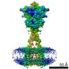 30853MC 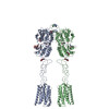 7dtuC 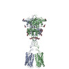 7dtvC 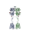 7dtwC M: map data used to model this data C: citing same article ( |
|---|---|
| Similar structure data |

-Links
 PDBj
PDBj


- Assembly
| Deposited unit | 
|
|---|---|
| 1 |
|
-Components
| #1: Protein | Mass: 123520.828 Da / Num. of mol.: 2 Source method: isolated from a genetically manipulated source Source: (gene. exp.) Homo sapiens (human) / Gene: CASR, GPRC2A, PCAR1 / Production host:   Spodoptera frugiperda (fall armyworm) / References: UniProt: P41180 Spodoptera frugiperda (fall armyworm) / References: UniProt: P41180#2: Polysaccharide | Source method: isolated from a genetically manipulated source #3: Polysaccharide |   Source method: isolated from a genetically manipulated source Details: oligosaccharide / References: triacetyl-beta-chitotriose #4: Chemical | ChemComp-CA /   Mass: 40.078 Da / Num. of mol.: 4 / Source method: obtained synthetically / Formula: Ca / Feature type: SUBJECT OF INVESTIGATION Mass: 40.078 Da / Num. of mol.: 4 / Source method: obtained synthetically / Formula: Ca / Feature type: SUBJECT OF INVESTIGATION#5: Sugar | ChemComp-NAG /   Type: D-saccharide, beta linking / Mass: 221.208 Da / Num. of mol.: 8 / Source method: obtained synthetically / Formula: C8H15NO6 Type: D-saccharide, beta linking / Mass: 221.208 Da / Num. of mol.: 8 / Source method: obtained synthetically / Formula: C8H15NO6Has ligand of interest | Y | Has protein modification | Y | |
|---|
-Experimental details
-Experiment
| Experiment | Method: ELECTRON MICROSCOPY |
|---|---|
| EM experiment | Aggregation state: PARTICLE / 3D reconstruction method: single particle reconstruction |
- Sample preparation
| Component | Name: Full-lenth Human Calcium-Sensing Receptor bound with 20 mM calcium ions Type: COMPLEX / Entity ID: #1 / Source: RECOMBINANT |
|---|---|
| Source (natural) | Organism: Homo sapiens (human) |
| Source (recombinant) | Organism: Spodoptera frugiperda (fall armyworm) |
| Buffer solution | pH: 7.5 |
| Specimen | Embedding applied: NO / Shadowing applied: NO / Staining applied: NO / Vitrification applied: YES |
| Vitrification | Cryogen name: ETHANE |
- Electron microscopy imaging
| Experimental equipment |  Model: Titan Krios / Image courtesy: FEI Company |
|---|---|
| Microscopy | Model: FEI TITAN KRIOS |
| Electron gun | Electron source:  FIELD EMISSION GUN / Accelerating voltage: 300 kV / Illumination mode: SPOT SCAN FIELD EMISSION GUN / Accelerating voltage: 300 kV / Illumination mode: SPOT SCAN |
| Electron lens | Mode: DIFFRACTION |
| Image recording | Electron dose: 62 e/Å2 / Detector mode: COUNTING / Film or detector model: GATAN K2 SUMMIT (4k x 4k) |
- Processing
| Software |
| ||||||||||||||||||||||||
|---|---|---|---|---|---|---|---|---|---|---|---|---|---|---|---|---|---|---|---|---|---|---|---|---|---|
| CTF correction | Type: PHASE FLIPPING AND AMPLITUDE CORRECTION | ||||||||||||||||||||||||
| 3D reconstruction | Resolution: 3.8 Å / Resolution method: FSC 0.143 CUT-OFF / Num. of particles: 315050 / Symmetry type: POINT | ||||||||||||||||||||||||
| Refinement | Stereochemistry target values: CDL v1.2 | ||||||||||||||||||||||||
| Refine LS restraints |
|
