 Movie
Movie Controller


+ Open data

- Basic information
| Entry | Database: PDB / ID: 7d5i | |||||||||||||||||||||||||||
|---|---|---|---|---|---|---|---|---|---|---|---|---|---|---|---|---|---|---|---|---|---|---|---|---|---|---|---|---|
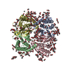
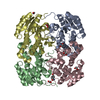
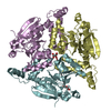
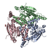
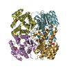
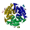
| Title | Structure of Mycobacterium smegmatis bd complex in the apo-form. | |||||||||||||||||||||||||||
 Components Components |
| |||||||||||||||||||||||||||
 Keywords Keywords | OXIDOREDUCTASE / Mycobacterium smegmatis / electron transfer chain | |||||||||||||||||||||||||||
| Function / homology |  Function and homology information Function and homology informationOxidoreductases; Acting on diphenols and related substances as donors; With oxygen as acceptor / cytochrome complex / alkaline phosphatase / alkaline phosphatase activity / aerobic electron transport chain / oxidoreductase activity, acting on diphenols and related substances as donors, oxygen as acceptor / electron transfer activity / heme binding / metal ion binding / plasma membrane Similarity search - Function | |||||||||||||||||||||||||||
| Biological species |  Mycolicibacterium smegmatis MC2 155 (bacteria) Mycolicibacterium smegmatis MC2 155 (bacteria) | |||||||||||||||||||||||||||
| Method | ELECTRON MICROSCOPY / single particle reconstruction / cryo EM / Resolution: 2.79 Å | |||||||||||||||||||||||||||
 Authors Authors | Wang, W. / Gong, H. / Gao, Y. / Zhou, X. / Rao, Z. | |||||||||||||||||||||||||||
| Funding support |  China, 3items China, 3items
| |||||||||||||||||||||||||||
 Citation Citation | Journal: Nat Commun / Year: 2021 Title: Cryo-EM structure of mycobacterial cytochrome bd reveals two oxygen access channels. Authors: Weiwei Wang / Yan Gao / Yanting Tang / Xiaoting Zhou / Yuezheng Lai / Shan Zhou / Yuying Zhang / Xiuna Yang / Fengjiang Liu / Luke W Guddat / Quan Wang / Zihe Rao / Hongri Gong /  Abstract: Cytochromes bd are ubiquitous amongst prokaryotes including many human-pathogenic bacteria. Such complexes are targets for the development of antimicrobial drugs. However, an understanding of the ...Cytochromes bd are ubiquitous amongst prokaryotes including many human-pathogenic bacteria. Such complexes are targets for the development of antimicrobial drugs. However, an understanding of the relationship between the structure and functional mechanisms of these oxidases is incomplete. Here, we have determined the 2.8 Å structure of Mycobacterium smegmatis cytochrome bd by single-particle cryo-electron microscopy. This bd oxidase consists of two subunits CydA and CydB, that adopt a pseudo two-fold symmetrical arrangement. The structural topology of its Q-loop domain, whose function is to bind the substrate, quinol, is significantly different compared to the C-terminal region reported for cytochromes bd from Geobacillus thermodenitrificans (G. th) and Escherichia coli (E. coli). In addition, we have identified two potential oxygen access channels in the structure and shown that similar tunnels also exist in G. th and E. coli cytochromes bd. This study provides insights to develop a framework for the rational design of antituberculosis compounds that block the oxygen access channels of this oxidase. | |||||||||||||||||||||||||||
| History |
|
- Structure visualization
| Movie |
Movie viewer |
|---|---|
| Structure viewer | Molecule: MolmilJmol/JSmol |
- Downloads & links
-Download
| PDBx/mmCIF format | 7d5i.cif.gz | 152.2 KB | Display | PDBx/mmCIF format |
|---|---|---|---|---|
| PDB format | pdb7d5i.ent.gz | 115.3 KB | Display | PDB format |
| PDBx/mmJSON format | 7d5i.json.gz | Tree view | PDBx/mmJSON format | |
| Others |  Other downloads Other downloads |
-Validation report
| Arichive directory | https://data.pdbj.org/pub/pdb/validation_reports/d5/7d5iftp://data.pdbj.org/pub/pdb/validation_reports/d5/7d5i | HTTPS FTP |
|---|
-Related structure data
| Related structure data |  30582MC M: map data used to model this data C: citing same article ( |
|---|---|
| Similar structure data |


-Links
 PDBj
PDBj
- Assembly
| Deposited unit | 
|
|---|---|
| 1 |
|
-Components
| #1: Protein | Mass: 53997.961 Da / Num. of mol.: 1 Source method: isolated from a genetically manipulated source Source: (gene. exp.) Mycolicibacterium smegmatis MC2 155 (bacteria)Strain: MC2 155 / Gene: MSMEG_3233 / Production host: References: UniProt: A0QXA8, Oxidoreductases; Acting on diphenols and related substances as donors; With oxygen as acceptor | ||||||
|---|---|---|---|---|---|---|---|
| #2: Protein | Mass: 39352.660 Da / Num. of mol.: 1 Source method: isolated from a genetically manipulated source Source: (gene. exp.) Mycolicibacterium smegmatis MC2 155 (bacteria)Strain: MC2 155 / Gene: cydB, MSMEI_3150 / Production host: | ||||||
| #3: Chemical |   Mass: 618.503 Da / Num. of mol.: 2 / Source method: obtained synthetically / Formula: C34H34FeN4O4 / Feature type: SUBJECT OF INVESTIGATION Mass: 618.503 Da / Num. of mol.: 2 / Source method: obtained synthetically / Formula: C34H34FeN4O4 / Feature type: SUBJECT OF INVESTIGATION#4: Chemical | ChemComp-HDD / |   Mass: 632.487 Da / Num. of mol.: 1 / Source method: obtained synthetically / Formula: C34H32FeN4O5 / Feature type: SUBJECT OF INVESTIGATION Mass: 632.487 Da / Num. of mol.: 1 / Source method: obtained synthetically / Formula: C34H32FeN4O5 / Feature type: SUBJECT OF INVESTIGATIONHas ligand of interest | Y | Has protein modification | N | |
-Experimental details
-Experiment
| Experiment | Method: ELECTRON MICROSCOPY |
|---|---|
| EM experiment | Aggregation state: PARTICLE / 3D reconstruction method: single particle reconstruction |
- Sample preparation
| Component | Name: Cryo-EM structure of Mycobacterium smegmatis bd complex Type: COMPLEX / Entity ID: #1-#2 / Source: RECOMBINANT |
|---|---|
| Source (natural) | Organism: Mycolicibacterium smegmatis MC2 155 (bacteria) |
| Source (recombinant) | Organism: |
| Buffer solution | pH: 7 |
| Specimen | Embedding applied: NO / Shadowing applied: NO / Staining applied: NO / Vitrification applied: YES |
| Vitrification | Instrument: FEI VITROBOT MARK II / Cryogen name: ETHANE / Humidity: 100 % / Chamber temperature: 281 K |
- Electron microscopy imaging
| Experimental equipment |  Model: Titan Krios / Image courtesy: FEI Company |
|---|---|
| Microscopy | Model: FEI TITAN KRIOS |
| Electron gun | Electron source:  FIELD EMISSION GUN / Accelerating voltage: 300 kV / Illumination mode: SPOT SCAN FIELD EMISSION GUN / Accelerating voltage: 300 kV / Illumination mode: SPOT SCAN |
| Electron lens | Mode: BRIGHT FIELD / Cs: 2.7 mm / C2 aperture diameter: 70 µm |
| Image recording | Average exposure time: 2 sec. / Electron dose: 50 e/Å2 / Film or detector model: GATAN K3 (6k x 4k) |
- Processing
| Software | Name: PHENIX / Version: 1.16_3549: / Classification: refinement | ||||||||||||||||||||||||
|---|---|---|---|---|---|---|---|---|---|---|---|---|---|---|---|---|---|---|---|---|---|---|---|---|---|
| EM software |
| ||||||||||||||||||||||||
| CTF correction | Type: PHASE FLIPPING AND AMPLITUDE CORRECTION | ||||||||||||||||||||||||
| 3D reconstruction | Resolution: 2.79 Å / Resolution method: FSC 0.143 CUT-OFF / Num. of particles: 270938 / Details: The software used for reconstruction is cryoSPARC. / Symmetry type: POINT | ||||||||||||||||||||||||
| Refine LS restraints |
|
