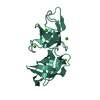
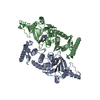
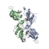
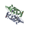
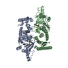
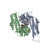
Journal: Nat Commun / Year: 2021 Title: Self-association of MreC as a regulatory signal in bacterial cell wall elongation. Authors: Alexandre Martins / Carlos Contreras-Martel / Manon Janet-Maitre / Mayara M Miyachiro / Leandro F Estrozi / Daniel Maragno Trindade / Caíque C Malospirito / Fernanda Rodrigues-Costa / ...Authors: Alexandre Martins / Carlos Contreras-Martel / Manon Janet-Maitre / Mayara M Miyachiro / Leandro F Estrozi / Daniel Maragno Trindade / Caíque C Malospirito / Fernanda Rodrigues-Costa / Lionel Imbert / Viviana Job / Guy Schoehn / Ina Attrée / Andréa Dessen / Abstract: The elongasome, or Rod system, is a protein complex that controls cell wall formation in rod-shaped bacteria. MreC is a membrane-associated elongasome component that co-localizes with the ...The elongasome, or Rod system, is a protein complex that controls cell wall formation in rod-shaped bacteria. MreC is a membrane-associated elongasome component that co-localizes with the cytoskeletal element MreB and regulates the activity of cell wall biosynthesis enzymes, in a process that may be dependent on MreC self-association. Here, we use electron cryo-microscopy and X-ray crystallography to determine the structure of a self-associated form of MreC from Pseudomonas aeruginosa in atomic detail. MreC monomers interact in head-to-tail fashion. Longitudinal and lateral interfaces are essential for oligomerization in vitro, and a phylogenetic analysis of proteobacterial MreC sequences indicates the prevalence of the identified interfaces. Our results are consistent with a model where MreC's ability to alternate between self-association and interaction with the cell wall biosynthesis machinery plays a key role in the regulation of elongasome activity.
A: Rod shape-determining protein MreC B: Rod shape-determining protein MreC C: Rod shape-determining protein MreC D: Rod shape-determining protein MreC
 Movie
Movie Controller


 Open data
 Basic information
 Components
 Keywords
 Function and homology information
 Pseudomonas aeruginosa (bacteria)
 Authors
 Brazil,
  France, 7items
  Citation
 Structure visualization
 Downloads & links
 Other downloads


 PDBj
PDBj Assembly
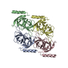
 Sample preparation
 Electron microscopy imaging
 FIELD EMISSION GUN / Accelerating voltage: 200 kV / Illumination mode: FLOOD BEAM
 Processing