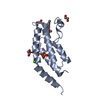
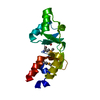
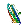
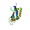
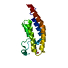
Cell/Rod shape-determining protein MreC, domain 1 / Rod shape-determining protein MreC / Cell/Rod shape-determining protein MreC, domain 2 / Rod shape-determining protein MreC, barrel core / regulation of cell shape / metal ion binding / plasma membrane / Cell shape-determining protein MreC
Function and homology information
Biological species
Pseudomonas aeruginosa PAO1 (bacteria)
Method
X-RAY DIFFRACTION / SYNCHROTRON / AB INITIO PHASING / Resolution: 1.471 Å
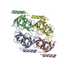
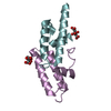
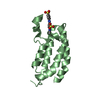
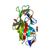
Journal: Nat Commun / Year: 2021 Title: Self-association of MreC as a regulatory signal in bacterial cell wall elongation. Authors: Alexandre Martins / Carlos Contreras-Martel / Manon Janet-Maitre / Mayara M Miyachiro / Leandro F Estrozi / Daniel Maragno Trindade / Caíque C Malospirito / Fernanda Rodrigues-Costa / ...Authors: Alexandre Martins / Carlos Contreras-Martel / Manon Janet-Maitre / Mayara M Miyachiro / Leandro F Estrozi / Daniel Maragno Trindade / Caíque C Malospirito / Fernanda Rodrigues-Costa / Lionel Imbert / Viviana Job / Guy Schoehn / Ina Attrée / Andréa Dessen / Abstract: The elongasome, or Rod system, is a protein complex that controls cell wall formation in rod-shaped bacteria. MreC is a membrane-associated elongasome component that co-localizes with the ...The elongasome, or Rod system, is a protein complex that controls cell wall formation in rod-shaped bacteria. MreC is a membrane-associated elongasome component that co-localizes with the cytoskeletal element MreB and regulates the activity of cell wall biosynthesis enzymes, in a process that may be dependent on MreC self-association. Here, we use electron cryo-microscopy and X-ray crystallography to determine the structure of a self-associated form of MreC from Pseudomonas aeruginosa in atomic detail. MreC monomers interact in head-to-tail fashion. Longitudinal and lateral interfaces are essential for oligomerization in vitro, and a phylogenetic analysis of proteobacterial MreC sequences indicates the prevalence of the identified interfaces. Our results are consistent with a model where MreC's ability to alternate between self-association and interaction with the cell wall biosynthesis machinery plays a key role in the regulation of elongasome activity.
Method to determine structure: AB INITIO PHASING / Resolution: 1.471→42.435 Å / Cor.coef. Fo:Fc: 0.966 / Cor.coef. Fo:Fc free: 0.955 / SU B: 4.582 / SU ML: 0.082 / Cross valid method: FREE R-VALUE / ESU R: 0.082 / ESU R Free: 0.083 Details: Hydrogens have been added in their riding positions
Rfactor
Num. reflection
% reflection
Rfree
0.2445
2036
7.473 %
Rwork
0.2168
25210
-
all
0.219
-
-
obs
-
27246
96.262 %
Solvent computation
Ion probe radii: 0.8 Å / Shrinkage radii: 0.8 Å / VDW probe radii: 1.2 Å / Solvent model: BABINET MODEL PLUS MASK
Displacement parameters
Biso mean: 39.496 Å2
Baniso -1
Baniso -2
Baniso -3
1-
0.658 Å2
0.329 Å2
-0 Å2
2-
-
0.658 Å2
0 Å2
3-
-
-
-2.133 Å2
Refinement step
Cycle: LAST / Resolution: 1.471→42.435 Å
Protein
Nucleic acid
Ligand
Solvent
Total
Num. atoms
1178
0
6
66
1250
Refine LS restraints
Refine-ID
Type
Dev ideal
Dev ideal target
Number
X-RAY DIFFRACTION
r_bond_refined_d
0.01
0.013
1198
X-RAY DIFFRACTION
r_bond_other_d
0.006
0.017
1162
X-RAY DIFFRACTION
r_angle_refined_deg
1.332
1.643
1631
X-RAY DIFFRACTION
r_angle_other_deg
1.292
1.573
2683
X-RAY DIFFRACTION
r_dihedral_angle_1_deg
6.701
5
154
X-RAY DIFFRACTION
r_dihedral_angle_2_deg
30.244
21.5
60
X-RAY DIFFRACTION
r_dihedral_angle_3_deg
12.028
15
198
X-RAY DIFFRACTION
r_dihedral_angle_4_deg
19.598
15
10
X-RAY DIFFRACTION
r_chiral_restr
0.067
0.2
157
X-RAY DIFFRACTION
r_gen_planes_refined
0.01
0.02
1343
X-RAY DIFFRACTION
r_gen_planes_other
0.001
0.02
239
X-RAY DIFFRACTION
r_nbd_refined
0.214
0.2
179
X-RAY DIFFRACTION
r_symmetry_nbd_other
0.198
0.2
1087
X-RAY DIFFRACTION
r_nbtor_refined
0.155
0.2
580
X-RAY DIFFRACTION
r_symmetry_nbtor_other
0.079
0.2
553
X-RAY DIFFRACTION
r_xyhbond_nbd_refined
0.143
0.2
42
X-RAY DIFFRACTION
r_symmetry_xyhbond_nbd_other
0.148
0.2
1
X-RAY DIFFRACTION
r_symmetry_nbd_refined
0.22
0.2
11
X-RAY DIFFRACTION
r_nbd_other
0.236
0.2
46
X-RAY DIFFRACTION
r_symmetry_xyhbond_nbd_refined
0.134
0.2
6
X-RAY DIFFRACTION
r_mcbond_it
1.351
1.724
619
X-RAY DIFFRACTION
r_mcbond_other
1.349
1.723
618
X-RAY DIFFRACTION
r_mcangle_it
1.766
2.582
772
X-RAY DIFFRACTION
r_mcangle_other
1.765
2.583
773
X-RAY DIFFRACTION
r_scbond_it
1.885
1.969
579
X-RAY DIFFRACTION
r_scbond_other
1.883
1.97
580
X-RAY DIFFRACTION
r_scangle_it
2.838
2.862
859
X-RAY DIFFRACTION
r_scangle_other
2.837
2.862
860
X-RAY DIFFRACTION
r_lrange_it
5.885
21.132
1188
X-RAY DIFFRACTION
r_lrange_other
5.87
20.916
1184
LS refinement shell
Refine-ID: X-RAY DIFFRACTION / Total num. of bins used: 20
Resolution (Å)
Rfactor Rfree
Num. reflection Rfree
Rfactor Rwork
Num. reflection Rwork
Rfactor all
Num. reflection all
Fsc free
Fsc work
% reflection obs (%)
WRfactor Rwork
1.471-1.509
0.453
84
0.467
1112
0.466
2031
0.321
0.223
58.8872
0.464
1.509-1.55
0.422
144
0.383
1647
0.386
2003
0.527
0.568
89.4159
0.375
1.55-1.595
0.369
172
0.341
1778
0.343
1954
0.566
0.612
99.7953
0.325
1.595-1.644
0.317
123
0.316
1781
0.316
1905
0.771
0.764
99.9475
0.301
1.644-1.698
0.344
134
0.288
1722
0.292
1856
0.788
0.806
100
0.264
1.698-1.757
0.296
126
0.264
1641
0.266
1767
0.83
0.855
100
0.232
1.757-1.823
0.251
129
0.241
1592
0.241
1722
0.898
0.897
99.9419
0.204
1.823-1.898
0.253
124
0.22
1538
0.223
1662
0.917
0.928
100
0.187
1.898-1.982
0.262
98
0.212
1482
0.215
1580
0.919
0.931
100
0.176
1.982-2.078
0.213
111
0.201
1429
0.202
1540
0.946
0.948
100
0.171
2.078-2.19
0.218
116
0.207
1330
0.208
1446
0.942
0.941
100
0.18
2.19-2.323
0.307
115
0.217
1291
0.224
1407
0.91
0.932
99.9289
0.192
2.323-2.483
0.246
103
0.221
1211
0.223
1314
0.911
0.933
100
0.2
2.483-2.681
0.256
89
0.235
1123
0.236
1212
0.923
0.931
100
0.219
2.681-2.936
0.24
86
0.227
1055
0.228
1141
0.928
0.933
100
0.228
2.936-3.28
0.251
83
0.224
946
0.226
1029
0.927
0.937
100
0.235
3.28-3.784
0.259
58
0.212
857
0.215
915
0.91
0.946
100
0.242
3.784-4.625
0.198
58
0.177
728
0.179
786
0.962
0.962
100
0.215
4.625-6.503
0.252
60
0.198
578
0.203
638
0.939
0.957
100
0.247
6.503-42.435
0.182
23
0.213
369
0.211
396
0.973
0.96
98.9899
0.257
Refinement TLS params.
Method: refined / Refine-ID: X-RAY DIFFRACTION
ID
L11 (°2)
L12 (°2)
L13 (°2)
L22 (°2)
L23 (°2)
L33 (°2)
S11 (Å °)
S12 (Å °)
S13 (Å °)
S21 (Å °)
S22 (Å °)
S23 (Å °)
S31 (Å °)
S32 (Å °)
S33 (Å °)
T11 (Å2)
T12 (Å2)
T13 (Å2)
T22 (Å2)
T23 (Å2)
T33 (Å2)
Origin x (Å)
Origin y (Å)
Origin z (Å)
1
20.6384
-1.4127
13.0893
13.3443
4.1067
27.3752
0.072
0.5859
-0.6629
0.2515
0.1983
-1.4749
0.0736
1.6459
-0.2703
0.2539
0.0233
0.0437
0.2215
0.0195
0.3337
5.2212
27.335
-2.3775
2
5.5315
1.4702
-0.7354
3.8211
0.4205
4.2142
0.029
-0.0583
0.0293
-0.0919
-0.0354
0.1175
0.2319
-0.2103
0.0064
0.2288
-0.0124
-0.0334
0.1409
-0.0129
0.2252
-15.7989
22.817
1.4011
3
28.3693
-4.0309
-4.2376
5.0504
5.1464
9.0224
-0.0214
-0.2004
-0.5966
0.1849
0.1951
-0.2351
0.4281
0.5115
-0.1736
0.3042
-0.0038
0.0093
0.2147
-0.0564
0.235
-3.2158
19.1562
-3.837
4
1.3274
0.1996
-0.5175
0.8509
-0.2851
2.8291
0.1221
-0.0626
-0.1372
-0.0497
-0.1197
-0.0709
0.0596
0.3044
-0.0024
0.1528
-0.0099
-0.0076
0.1436
-0.0014
0.2012
-4.5693
26.5282
7.2618
5
7.0684
2.0885
1.6869
5.3597
0.7725
7.3564
0.2234
0.0661
0.0676
0.0541
-0.0248
0.0799
-0.2232
-0.0792
-0.1985
0.2164
0.0026
0.0581
0.1641
-0.003
0.1978
-13.7609
33.7
14.162
6
2.4633
3.0969
-4.305
3.9101
-5.4552
7.6994
0.5894
-0.2451
0.1942
0.7514
-0.2121
0.2225
-1.1791
0.2037
-0.3773
0.4606
-0.1334
0.1673
0.7472
-0.2652
0.2524
-11.0993
40.4814
24.7947
7
5.1865
-2.2525
-0.1675
8.2238
-0.2501
4.3541
0.2948
-0.0404
0.2315
0.0998
-0.2713
0.1775
-0.6734
0.5175
-0.0235
0.2494
-0.1484
0.052
0.199
-0.0335
0.182
-4.9986
40.5683
15.5848
8
1.9943
-0.8346
0.1021
4.931
0.2362
4.9673
0.2515
-0.1087
-0.2608
0.2505
-0.1255
0.1811
-0.3951
0.515
-0.126
0.213
-0.0929
0.0286
0.2198
-0.0207
0.2013
-4.8178
35.6599
12.1059
9
2.7217
-0.1721
-2.2912
1.8632
-0.6151
9.0215
0.3241
-0.0195
0.1497
0.2194
-0.0333
0.2061
-0.9193
-0.4233
-0.2908
0.2658
-0.0254
0.0576
0.1433
-0.0324
0.2587
-15.6645
38.9103
18.0427
10
16.0908
-9.3143
1.6742
11.238
0.528
6.7049
-0.0434
0.8369
-0.1746
-0.5559
-0.3482
0.124
-0.4962
-0.0023
0.3917
0.2993
-0.0156
0.0059
0.1851
-0.0363
0.244
-2.9336
29.9247
-2.4384
Refinement TLS group
ID
Refine-ID
Refine TLS-ID
Selection
Auth asym-ID
Auth seq-ID
1
X-RAY DIFFRACTION
1
ALL
AAA
100 - 105
2
X-RAY DIFFRACTION
2
ALL
AAA
106 - 119
3
X-RAY DIFFRACTION
3
ALL
AAA
120 - 125
4
X-RAY DIFFRACTION
4
ALL
AAA
126 - 174
5
X-RAY DIFFRACTION
5
ALL
AAA
175 - 188
6
X-RAY DIFFRACTION
6
ALL
AAA
189 - 198
7
X-RAY DIFFRACTION
7
ALL
AAA
199 - 210
8
X-RAY DIFFRACTION
8
ALL
AAA
211 - 224
9
X-RAY DIFFRACTION
9
ALL
AAA
225 - 245
10
X-RAY DIFFRACTION
10
ALL
AAA
246 - 254
+
About Yorodumi
-
News
-
Feb 9, 2022. New format data for meta-information of EMDB entries
New format data for meta-information of EMDB entries
Version 3 of the EMDB header file is now the official format.
The previous official version 1.9 will be removed from the archive.
In the structure databanks used in Yorodumi, some data are registered as the other names, "COVID-19 virus" and "2019-nCoV". Here are the details of the virus and the list of structure data.
Jan 31, 2019. EMDB accession codes are about to change! (news from PDBe EMDB page)
EMDB accession codes are about to change! (news from PDBe EMDB page)
The allocation of 4 digits for EMDB accession codes will soon come to an end. Whilst these codes will remain in use, new EMDB accession codes will include an additional digit and will expand incrementally as the available range of codes is exhausted. The current 4-digit format prefixed with “EMD-” (i.e. EMD-XXXX) will advance to a 5-digit format (i.e. EMD-XXXXX), and so on. It is currently estimated that the 4-digit codes will be depleted around Spring 2019, at which point the 5-digit format will come into force.
The EM Navigator/Yorodumi systems omit the EMD- prefix.
Related info.:Q: What is EMD? / ID/Accession-code notation in Yorodumi/EM Navigator
Yorodumi is a browser for structure data from EMDB, PDB, SASBDB, etc.
This page is also the successor to EM Navigator detail page, and also detail information page/front-end page for Omokage search.
The word "yorodu" (or yorozu) is an old Japanese word meaning "ten thousand". "mi" (miru) is to see.
Related info.:EMDB / PDB / SASBDB / Comparison of 3 databanks / Yorodumi Search / Aug 31, 2016. New EM Navigator & Yorodumi / Yorodumi Papers / Jmol/JSmol / Function and homology information / Changes in new EM Navigator and Yorodumi
 Movie
Movie Controller


 Open data
 Basic information
 Components
 Keywords
 Function and homology information
 Pseudomonas aeruginosa PAO1 (bacteria)
 X-RAY DIFFRACTION /
  Authors
 France,
  Brazil, 3items
  Citation
 Structure visualization
 Downloads & links
 Other downloads

 PDBj
PDBj Assembly

 Mass: 24.305 Da / Num. of mol.: 1 / Source method: obtained synthetically / Formula: Mg
 Mass: 35.453 Da / Num. of mol.: 5 / Source method: obtained synthetically / Formula: Cl
 Mass: 18.015 Da / Num. of mol.: 66 / Source method: isolated from a natural source / Formula: H2O
 Sample preparation
 Processing