 ムービー
ムービー コントローラー


+ データを開く

- 基本情報
基本情報
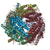
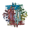
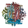
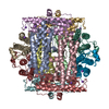
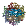
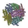
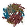
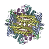
| 登録情報 | データベース: PDB / ID: 6zgl | ||||||
|---|---|---|---|---|---|---|---|
| タイトル | Structure of DPS determined by movement-free cryoEM with zero dose extrapolation | ||||||
 要素 要素 | DNA protection during starvation protein | ||||||
 キーワード キーワード | DNA BINDING PROTEIN / DNA-BINDING PROTEIN | ||||||
| 機能・相同性 |  機能・相同性情報 機能・相同性情報DnaA-Dps complex / 酸化還元酵素; 金属イオンを酸化する / oxidoreductase activity, acting on metal ions / nucleoid / chromosome condensation / response to starvation / response to stress / negative regulation of DNA-templated DNA replication initiation / ferric iron binding / intracellular iron ion homeostasis ...DnaA-Dps complex / 酸化還元酵素; 金属イオンを酸化する / oxidoreductase activity, acting on metal ions / nucleoid / chromosome condensation / response to starvation / response to stress / negative regulation of DNA-templated DNA replication initiation / ferric iron binding / intracellular iron ion homeostasis / DNA binding / identical protein binding / membrane / cytoplasm 類似検索 - 分子機能 | ||||||
| 生物種 |  | ||||||
| 手法 | 電子顕微鏡法 / 単粒子再構成法 / クライオ電子顕微鏡法 / 解像度: 1.9 Å | ||||||
 データ登録者 データ登録者 | Naydenova, K. / Russo, C.J. | ||||||
| 資金援助 |  英国, 1件 英国, 1件
| ||||||
 引用 引用 | ジャーナル: Science / 年: 2020 タイトル: Cryo-EM with sub-1 Å specimen movement. 著者: Katerina Naydenova / Peipei Jia / Christopher J Russo /  要旨: Most information loss in cryogenic electron microscopy (cryo-EM) stems from particle movement during imaging, which remains poorly understood. We show that this movement is caused by buckling and ...Most information loss in cryogenic electron microscopy (cryo-EM) stems from particle movement during imaging, which remains poorly understood. We show that this movement is caused by buckling and subsequent deformation of the suspended ice, with a threshold that depends directly on the shape of the frozen water layer set by the support foil. We describe a specimen support design that eliminates buckling and reduces electron beam-induced particle movement to less than 1 angstrom. The design allows precise foil tracking during imaging with high-speed detectors, thereby lessening demands on cryostage precision and stability. It includes a maximal density of holes, which increases throughput in automated cryo-EM without degrading data quality. Movement-free imaging allows extrapolation to a three-dimensional map of the specimen at zero electron exposure, before the onset of radiation damage. | ||||||
| 履歴 |
|
- 構造の表示
構造の表示
| ムービー |
ムービービューア |
|---|---|
| 構造ビューア | 分子: MolmilJmol/JSmol |
- ダウンロードとリンク
-ダウンロード
| PDBx/mmCIF形式 | 6zgl.cif.gz | 380.4 KB | 表示 | PDBx/mmCIF形式 |
|---|---|---|---|---|
| PDB形式 | pdb6zgl.ent.gz | 311.1 KB | 表示 | PDB形式 |
| PDBx/mmJSON形式 | 6zgl.json.gz | ツリー表示 | PDBx/mmJSON形式 | |
| その他 |  その他のダウンロード その他のダウンロード |
-検証レポート
| 文書・要旨 | 6zgl_validation.pdf.gz | 886.9 KB | 表示 | wwPDB検証レポート |
|---|---|---|---|---|
| 文書・詳細版 | 6zgl_full_validation.pdf.gz | 909 KB | 表示 | |
| XML形式データ | 6zgl_validation.xml.gz | 49.6 KB | 表示 | |
| CIF形式データ | 6zgl_validation.cif.gz | 71.2 KB | 表示 | |
| アーカイブディレクトリ | https://data.pdbj.org/pub/pdb/validation_reports/zg/6zglftp://data.pdbj.org/pub/pdb/validation_reports/zg/6zgl | HTTPS FTP |
-関連構造データ
| 関連構造データ | 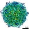 11210MC M: このデータのモデリングに利用したマップデータ C: 同じ文献を引用 ( |
|---|---|
| 類似構造データ | |
| 電子顕微鏡画像生データ | EMPIAR-10445 (タイトル: Movies of DPS in 260 nm gold foil holes, which eliminate specimen movement Data size: 1.5 TB Data #1: Unaligned multi-frame micrographs of DPS in 260 nm hole supports [micrographs - multiframe]) |

-リンク
 PDBj
PDBj


- 集合体
集合体
| 登録構造単位 | 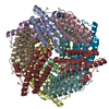
|
|---|---|
| 1 |
|
-要素
| #1: タンパク質 | 分子量: 18720.295 Da / 分子数: 12 / 由来タイプ: 組換発現 / 由来: (組換発現) #2: 水 | ChemComp-HOH / |  分子量: 18.015 Da / 分子数: 964 / 由来タイプ: 天然 / 式: H2O 分子量: 18.015 Da / 分子数: 964 / 由来タイプ: 天然 / 式: H2O |
|---|
-実験情報
-実験
| 実験 | 手法: 電子顕微鏡法 |
|---|---|
| EM実験 | 試料の集合状態: PARTICLE / 3次元再構成法: 単粒子再構成法 |
- 試料調製
試料調製
| 構成要素 | 名称: DNA protection during starvation protein (DPS) / タイプ: COMPLEX / Entity ID: #1 / 由来: RECOMBINANT |
|---|---|
| 分子量 | 実験値: NO |
| 由来(天然) | 生物種: |
| 由来(組換発現) | 生物種: |
| 緩衝液 | pH: 7.7 |
| 試料 | 包埋: NO / シャドウイング: NO / 染色: NO / 凍結: YES |
| 急速凍結 | 凍結剤: ETHANE |
- 電子顕微鏡撮影
| 実験機器 |  モデル: Titan Krios / 画像提供: FEI Company |
|---|---|
| 顕微鏡 | モデル: FEI TITAN KRIOS |
| 電子銃 | 電子線源:  FIELD EMISSION GUN / 加速電圧: 300 kV / 照射モード: FLOOD BEAM FIELD EMISSION GUN / 加速電圧: 300 kV / 照射モード: FLOOD BEAM |
| 電子レンズ | モード: BRIGHT FIELD |
| 撮影 | 電子線照射量: 35 e/Å2 フィルム・検出器のモデル: FEI FALCON IV (4k x 4k) |
- 解析
解析
| ソフトウェア | 名称: REFMAC / バージョン: 5.8.0256 / 分類: 精密化 | ||||||||||||||||||||||||||||||||||||||||||||||||||||||||||||||||||||||||||||||||||||||||||||||||||||||||||
|---|---|---|---|---|---|---|---|---|---|---|---|---|---|---|---|---|---|---|---|---|---|---|---|---|---|---|---|---|---|---|---|---|---|---|---|---|---|---|---|---|---|---|---|---|---|---|---|---|---|---|---|---|---|---|---|---|---|---|---|---|---|---|---|---|---|---|---|---|---|---|---|---|---|---|---|---|---|---|---|---|---|---|---|---|---|---|---|---|---|---|---|---|---|---|---|---|---|---|---|---|---|---|---|---|---|---|---|
| CTF補正 | タイプ: PHASE FLIPPING AND AMPLITUDE CORRECTION | ||||||||||||||||||||||||||||||||||||||||||||||||||||||||||||||||||||||||||||||||||||||||||||||||||||||||||
| 対称性 | 点対称性: T (正4面体型対称) | ||||||||||||||||||||||||||||||||||||||||||||||||||||||||||||||||||||||||||||||||||||||||||||||||||||||||||
| 3次元再構成 | 解像度: 1.9 Å / 解像度の算出法: FSC 0.143 CUT-OFF / 粒子像の数: 275623 / 対称性のタイプ: POINT | ||||||||||||||||||||||||||||||||||||||||||||||||||||||||||||||||||||||||||||||||||||||||||||||||||||||||||
| 精密化 | 解像度: 1.9→1.9 Å / Cor.coef. Fo:Fc: 0.764 / ESU R: 0.168 立体化学のターゲット値: MAXIMUM LIKELIHOOD WITH PHASES 詳細: HYDROGENS HAVE BEEN ADDED IN THE RIDING POSITIONS
| ||||||||||||||||||||||||||||||||||||||||||||||||||||||||||||||||||||||||||||||||||||||||||||||||||||||||||
| 溶媒の処理 | イオンプローブ半径: 0.8 Å / 減衰半径: 0.8 Å / VDWプローブ半径: 1.2 Å / 溶媒モデル: MASK | ||||||||||||||||||||||||||||||||||||||||||||||||||||||||||||||||||||||||||||||||||||||||||||||||||||||||||
| 原子変位パラメータ | Biso mean: 19.636 Å2
| ||||||||||||||||||||||||||||||||||||||||||||||||||||||||||||||||||||||||||||||||||||||||||||||||||||||||||
| 精密化ステップ | サイクル: 1 / 合計: 15616 | ||||||||||||||||||||||||||||||||||||||||||||||||||||||||||||||||||||||||||||||||||||||||||||||||||||||||||
| 拘束条件 |
|