Spanish Ministry of Science, Innovation, and Universities
BFU 2014-54181
Spain
Spanish Ministry of Science, Innovation, and Universities
SEV-2013-0347
Spain
Spanish Ministry of Science, Innovation, and Universities
BES-2015-073615
Spain
Citation
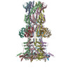
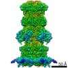
Journal: Proc Natl Acad Sci U S A / Year: 2021 Title: Assisted assembly of bacteriophage T7 core components for genome translocation across the bacterial envelope. Authors: Mar Pérez-Ruiz / Mar Pulido-Cid / Juan Román Luque-Ortega / José María Valpuesta / Ana Cuervo / José L Carrascosa / Abstract: In most bacteriophages, genome transport across bacterial envelopes is carried out by the tail machinery. In viruses of the family, in which the tail is not long enough to traverse the bacterial ...In most bacteriophages, genome transport across bacterial envelopes is carried out by the tail machinery. In viruses of the family, in which the tail is not long enough to traverse the bacterial wall, it has been postulated that viral core proteins assembled inside the viral head are translocated and reassembled into a tube within the periplasm that extends the tail channel. Bacteriophage T7 infects , and despite extensive studies, the precise mechanism by which its genome is translocated remains unknown. Using cryo-electron microscopy, we have resolved the structure of two different assemblies of the T7 DNA translocation complex composed of the core proteins gp15 and gp16. Gp15 alone forms a partially folded hexamer, which is further assembled upon interaction with gp16 into a tubular structure, forming a channel that could allow DNA passage. The structure of the gp15-gp16 complex also shows the location within gp16 of a canonical transglycosylase motif involved in the degradation of the bacterial peptidoglycan layer. This complex docks well in the tail extension structure found in the periplasm of T7-infected bacteria and matches the sixfold symmetry of the phage tail. In such cases, gp15 and gp16 that are initially present in the T7 capsid eightfold-symmetric core would change their oligomeric state upon reassembly in the periplasm. Altogether, these results allow us to propose a model for the assembly of the core translocation complex in the periplasm, which furthers understanding of the molecular mechanism involved in the release of T7 viral DNA into the bacterial cytoplasm.
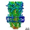
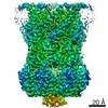
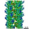
A: Internal virion protein gp15 B: Internal virion protein gp15 C: Internal virion protein gp15 D: Internal virion protein gp15 E: Internal virion protein gp15 F: Internal virion protein gp15
Average exposure time: 35 sec. / Electron dose: 30 e/Å2 / Detector mode: COUNTING / Film or detector model: FEI FALCON III (4k x 4k) / Num. of grids imaged: 1 / Num. of real images: 2470
Image scans
Movie frames/image: 32
-
Processing
EM software
ID
Name
Category
1
Gautomatch
particleselection
2
EPU
imageacquisition
4
CTFFIND
CTFcorrection
7
Coot
modelfitting
9
PHENIX
modelrefinement
10
RELION
initialEulerassignment
11
RELION
finalEulerassignment
12
RELION
classification
13
RELION
3Dreconstruction
CTF correction
Type: PHASE FLIPPING AND AMPLITUDE CORRECTION
Symmetry
Point symmetry: C6 (6 fold cyclic)
3D reconstruction
Resolution: 3.6 Å / Resolution method: FSC 0.143 CUT-OFF / Num. of particles: 50980 / Algorithm: FOURIER SPACE / Num. of class averages: 1 / Symmetry type: POINT
Atomic model building
Protocol: AB INITIO MODEL / Space: REAL
+
About Yorodumi
-
News
-
Feb 9, 2022. New format data for meta-information of EMDB entries
New format data for meta-information of EMDB entries
Version 3 of the EMDB header file is now the official format.
The previous official version 1.9 will be removed from the archive.
In the structure databanks used in Yorodumi, some data are registered as the other names, "COVID-19 virus" and "2019-nCoV". Here are the details of the virus and the list of structure data.
Jan 31, 2019. EMDB accession codes are about to change! (news from PDBe EMDB page)
EMDB accession codes are about to change! (news from PDBe EMDB page)
The allocation of 4 digits for EMDB accession codes will soon come to an end. Whilst these codes will remain in use, new EMDB accession codes will include an additional digit and will expand incrementally as the available range of codes is exhausted. The current 4-digit format prefixed with “EMD-” (i.e. EMD-XXXX) will advance to a 5-digit format (i.e. EMD-XXXXX), and so on. It is currently estimated that the 4-digit codes will be depleted around Spring 2019, at which point the 5-digit format will come into force.
The EM Navigator/Yorodumi systems omit the EMD- prefix.
Related info.:Q: What is EMD? / ID/Accession-code notation in Yorodumi/EM Navigator
Yorodumi is a browser for structure data from EMDB, PDB, SASBDB, etc.
This page is also the successor to EM Navigator detail page, and also detail information page/front-end page for Omokage search.
The word "yorodu" (or yorozu) is an old Japanese word meaning "ten thousand". "mi" (miru) is to see.
Related info.:EMDB / PDB / SASBDB / Comparison of 3 databanks / Yorodumi Search / Aug 31, 2016. New EM Navigator & Yorodumi / Yorodumi Papers / Jmol/JSmol / Function and homology information / Changes in new EM Navigator and Yorodumi
 Movie
Movie Controller

 Yorodumi
 Open data
 Basic information
 Components
 Keywords
 Function and homology information
 Escherichia phage T7 (virus)
 Authors
 Spain, 3items
  Citation
 Structure visualization
 Downloads & links
 Other downloads
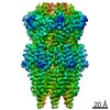


 PDBj
PDBj Assembly
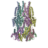

 Sample preparation
 Electron microscopy imaging
 FIELD EMISSION GUN / Accelerating voltage: 200 kV / Illumination mode: FLOOD BEAM
 Processing