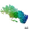
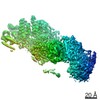
 Movie
Movie Controller

[English] 日本語
 Yorodumi
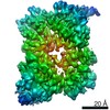
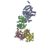
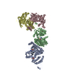
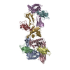
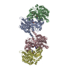
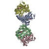
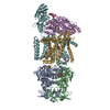
- PDB-6xqn: Structure of a mitochondrial calcium uniporter holocomplex (MICU1... -
+ Open data

- Basic information
| Entry | Database: PDB / ID: 6xqn | |||||||||
|---|---|---|---|---|---|---|---|---|---|---|
| Title | Structure of a mitochondrial calcium uniporter holocomplex (MICU1, MICU2, MCU, EMRE) in low Ca2+ | |||||||||
 Components Components |
| |||||||||
 Keywords Keywords | TRANSPORT PROTEIN/CALCIUM BINDING PROTEIN / ion channel / calcium channel / mitochondrial calcium uniporter / MCU / EMRE / mitochondria / TRANSPORT PROTEIN-CALCIUM BINDING PROTEIN complex | |||||||||
| Function / homology |  Function and homology information Function and homology informationmitochondrial crista junction / negative regulation of mitochondrial calcium ion concentration / regulation of cellular hyperosmotic salinity response / positive regulation of cristae formation / uniporter activity / uniplex complex / Processing of SMDT1 / positive regulation of mitochondrial calcium ion concentration / Mitochondrial calcium ion transport / mitochondrial calcium ion transmembrane transport ...mitochondrial crista junction / negative regulation of mitochondrial calcium ion concentration / regulation of cellular hyperosmotic salinity response / positive regulation of cristae formation / uniporter activity / uniplex complex / Processing of SMDT1 / positive regulation of mitochondrial calcium ion concentration / Mitochondrial calcium ion transport / mitochondrial calcium ion transmembrane transport / mitochondrial calcium ion homeostasis / calcium import into the mitochondrion / calcium ion sensor activity / calcium ion import / cellular response to calcium ion starvation / calcium channel inhibitor activity / calcium channel complex / Mitochondrial protein degradation / cellular response to calcium ion / calcium channel regulator activity / defense response / protein homooligomerization / mitochondrial intermembrane space / mitochondrial membrane / calcium channel activity / mitochondrial inner membrane / protein heterodimerization activity / calcium ion binding / mitochondrion / metal ion binding / identical protein binding Similarity search - Function | |||||||||
| Biological species |   Homo sapiens (human) Homo sapiens (human) | |||||||||
| Method | ELECTRON MICROSCOPY / single particle reconstruction / cryo EM / Resolution: 3.3 Å | |||||||||
 Authors Authors | Long, S.B. / Wang, C. / Baradaran, R. / Jacewicz, A. / Delgado, B. | |||||||||
| Funding support |  United States, 2items United States, 2items
| |||||||||
 Citation Citation | Journal: Elife / Year: 2020 Title: Structures reveal gatekeeping of the mitochondrial Ca uniporter by MICU1-MICU2. Authors: Chongyuan Wang / Agata Jacewicz / Bryce D Delgado / Rozbeh Baradaran / Stephen Barstow Long / Abstract: The mitochondrial calcium uniporter is a Ca-gated ion channel complex that controls mitochondrial Ca entry and regulates cell metabolism. MCU and EMRE form the channel while Ca-dependent regulation ...The mitochondrial calcium uniporter is a Ca-gated ion channel complex that controls mitochondrial Ca entry and regulates cell metabolism. MCU and EMRE form the channel while Ca-dependent regulation is conferred by MICU1 and MICU2 through an enigmatic process. We present a cryo-EM structure of an MCU-EMRE-MICU1-MICU2 holocomplex comprising MCU and EMRE subunits from the beetle Tribolium castaneum in complex with a human MICU1-MICU2 heterodimer at 3.3 Å resolution. With analogy to how neuronal channels are blocked by protein toxins, a uniporter interaction domain on MICU1 binds to a channel receptor site comprising MCU and EMRE subunits to inhibit ion flow under resting Ca conditions. A Ca-bound structure of MICU1-MICU2 at 3.1 Å resolution indicates how Ca-dependent changes enable dynamic response to cytosolic Ca signals. | |||||||||
| History |
|
- Structure visualization
| Movie |
Movie viewer |
|---|---|
| Structure viewer | Molecule: MolmilJmol/JSmol |
- Downloads & links
-Download
| PDBx/mmCIF format | 6xqn.cif.gz | 237.5 KB | Display | PDBx/mmCIF format |
|---|---|---|---|---|
| PDB format | pdb6xqn.ent.gz | 178 KB | Display | PDB format |
| PDBx/mmJSON format | 6xqn.json.gz | Tree view | PDBx/mmJSON format | |
| Others |  Other downloads Other downloads |
-Validation report
| Arichive directory | https://data.pdbj.org/pub/pdb/validation_reports/xq/6xqnftp://data.pdbj.org/pub/pdb/validation_reports/xq/6xqn | HTTPS FTP |
|---|
-Related structure data
| Related structure data | 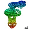 22290MC 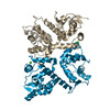 6xqoC M: map data used to model this data C: citing same article ( |
|---|---|
| Similar structure data |


-Links
 PDBj
PDBj

- Assembly
| Deposited unit | 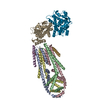
|
|---|---|
| 1 |
|
-Components
| #1: Protein | Mass: 7372.190 Da / Num. of mol.: 3 Source method: isolated from a genetically manipulated source Source: (gene. exp.) Homo sapiens (human) / References: UniProt: D6X268#2: Protein | Mass: 23634.100 Da / Num. of mol.: 4 Source method: isolated from a genetically manipulated source Source: (gene. exp.) Homo sapiens (human) / References: UniProt: D6WIX5#3: Protein | | Mass: 45422.883 Da / Num. of mol.: 1 Source method: isolated from a genetically manipulated source Source: (gene. exp.) Homo sapiens (human) / Gene: MICU1, CALC, CBARA1 / Production host: Homo sapiens (human) / References: UniProt: Q9BPX6#4: Protein | | Mass: 44980.609 Da / Num. of mol.: 1 Source method: isolated from a genetically manipulated source Source: (gene. exp.) Homo sapiens (human) / Gene: MICU2, EFHA1 / Production host: Homo sapiens (human) / References: UniProt: Q8IYU8#5: Chemical | ChemComp-CA / |   Mass: 40.078 Da / Num. of mol.: 1 / Source method: obtained synthetically / Formula: Ca Mass: 40.078 Da / Num. of mol.: 1 / Source method: obtained synthetically / Formula: CaHas ligand of interest | N | |
|---|
-Experimental details
-Experiment
| Experiment | Method: ELECTRON MICROSCOPY |
|---|---|
| EM experiment | Aggregation state: PARTICLE / 3D reconstruction method: single particle reconstruction |
- Sample preparation
| Component |
| ||||||||||||||||||||||||||||||
|---|---|---|---|---|---|---|---|---|---|---|---|---|---|---|---|---|---|---|---|---|---|---|---|---|---|---|---|---|---|---|---|
| Molecular weight | Units: MEGADALTONS / Experimental value: NO | ||||||||||||||||||||||||||||||
| Source (natural) |
| ||||||||||||||||||||||||||||||
| Source (recombinant) |
| ||||||||||||||||||||||||||||||
| Buffer solution | pH: 7.5 | ||||||||||||||||||||||||||||||
| Buffer component |
| ||||||||||||||||||||||||||||||
| Specimen | Conc.: 1 mg/ml / Embedding applied: NO / Shadowing applied: NO / Staining applied: NO / Vitrification applied: YES / Details: Monodisperse sample | ||||||||||||||||||||||||||||||
| Specimen support | Grid material: GOLD / Grid mesh size: 400 divisions/in. / Grid type: Quantifoil R1.2/1.3 | ||||||||||||||||||||||||||||||
| Vitrification | Instrument: FEI VITROBOT MARK IV / Cryogen name: ETHANE / Humidity: 100 % / Chamber temperature: 298 K / Details: 2 second blot, blot force of 0 |
- Electron microscopy imaging
| Experimental equipment |  Model: Titan Krios / Image courtesy: FEI Company |
|---|---|
| Microscopy | Model: FEI TITAN KRIOS |
| Electron gun | Electron source:  FIELD EMISSION GUN / Accelerating voltage: 300 kV / Illumination mode: FLOOD BEAM FIELD EMISSION GUN / Accelerating voltage: 300 kV / Illumination mode: FLOOD BEAM |
| Electron lens | Mode: BRIGHT FIELD / Nominal magnification: 22500 X / Nominal defocus max: -3000 nm / Nominal defocus min: -1000 nm / Alignment procedure: BASIC |
| Specimen holder | Cryogen: NITROGEN / Specimen holder model: FEI TITAN KRIOS AUTOGRID HOLDER |
| Image recording | Average exposure time: 4 sec. / Electron dose: 71 e/Å2 / Detector mode: SUPER-RESOLUTION / Film or detector model: GATAN K3 BIOQUANTUM (6k x 4k) / Num. of grids imaged: 5 / Num. of real images: 21115 |
| Image scans | Movie frames/image: 40 / Used frames/image: 1-40 |
- Processing
| EM software |
| ||||||||||||||||||||||||||||||||||||||||
|---|---|---|---|---|---|---|---|---|---|---|---|---|---|---|---|---|---|---|---|---|---|---|---|---|---|---|---|---|---|---|---|---|---|---|---|---|---|---|---|---|---|
| CTF correction | Type: PHASE FLIPPING AND AMPLITUDE CORRECTION | ||||||||||||||||||||||||||||||||||||||||
| Particle selection | Num. of particles selected: 17440131 | ||||||||||||||||||||||||||||||||||||||||
| Symmetry | Point symmetry: C2 (2 fold cyclic) | ||||||||||||||||||||||||||||||||||||||||
| 3D reconstruction | Resolution: 3.3 Å / Resolution method: FSC 0.143 CUT-OFF / Num. of particles: 350160 / Algorithm: FOURIER SPACE / Symmetry type: POINT | ||||||||||||||||||||||||||||||||||||||||
| Atomic model building | B value: 124 / Protocol: AB INITIO MODEL / Space: REAL / Target criteria: Correlation coefficient |
