ムービー
ムービー コントローラー


+ データを開く

- 基本情報
基本情報
| 登録情報 | データベース: PDB / ID: 6xe9 | ||||||||||||
|---|---|---|---|---|---|---|---|---|---|---|---|---|---|
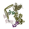
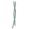
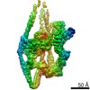
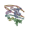
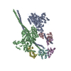
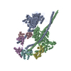
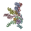
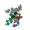
| タイトル | 10S myosin II (smooth muscle) | ||||||||||||
 要素 要素 |
| ||||||||||||
 キーワード キーワード | CONTRACTILE PROTEIN / motor protein / auto-inhibited form / 10 S conformation | ||||||||||||
| 機能・相同性 |  機能・相同性情報 機能・相同性情報myosin filament / actomyosin structure organization / myosin II complex / structural constituent of muscle / microfilament motor activity / myofibril / supramolecular fiber organization / ADP binding / actin filament binding / calmodulin binding ...myosin filament / actomyosin structure organization / myosin II complex / structural constituent of muscle / microfilament motor activity / myofibril / supramolecular fiber organization / ADP binding / actin filament binding / calmodulin binding / calcium ion binding / ATP binding / cytosol 類似検索 - 分子機能 | ||||||||||||
| 生物種 |  | ||||||||||||
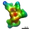
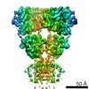
| 手法 | 電子顕微鏡法 / 単粒子再構成法 / クライオ電子顕微鏡法 / 解像度: 4.3 Å | ||||||||||||
 データ登録者 データ登録者 | Tiwari, P. / Craig, R. / Padron, R. | ||||||||||||
| 資金援助 |  米国, 3件 米国, 3件
| ||||||||||||
 引用 引用 | ジャーナル: Nature / 年: 2020 タイトル: Cryo-EM structure of the inhibited (10S) form of myosin II. 著者: Shixin Yang / Prince Tiwari / Kyoung Hwan Lee / Osamu Sato / Mitsuo Ikebe / Raúl Padrón / Roger Craig / 要旨: Myosin II is the motor protein that enables muscle cells to contract and nonmuscle cells to move and change shape. The molecule has two identical heads attached to an elongated tail, and can exist in ...Myosin II is the motor protein that enables muscle cells to contract and nonmuscle cells to move and change shape. The molecule has two identical heads attached to an elongated tail, and can exist in two conformations: 10S and 6S, named for their sedimentation coefficients. The 6S conformation has an extended tail and assembles into polymeric filaments, which pull on actin filaments to generate force and motion. In 10S myosin, the tail is folded into three segments and the heads bend back and interact with each other and the tail, creating a compact conformation in which ATPase activity, actin activation and filament assembly are all highly inhibited. This switched-off structure appears to function as a key energy-conserving storage molecule in muscle and nonmuscle cells, which can be activated to form functional filaments as needed-but the mechanism of its inhibition is not understood. Here we have solved the structure of smooth muscle 10S myosin by cryo-electron microscopy with sufficient resolution to enable improved understanding of the function of the head and tail regions of the molecule and of the key intramolecular contacts that cause inhibition. Our results suggest an atomic model for the off state of myosin II, for its activation and unfolding by phosphorylation, and for understanding the clustering of disease-causing mutations near sites of intramolecular interaction. | ||||||||||||
| 履歴 |
|
- 構造の表示
構造の表示
| ムービー |
ムービービューア |
|---|---|
| 構造ビューア | 分子: MolmilJmol/JSmol |
- ダウンロードとリンク
-ダウンロード
| PDBx/mmCIF形式 | 6xe9.cif.gz | 543 KB | 表示 | PDBx/mmCIF形式 |
|---|---|---|---|---|
| PDB形式 | pdb6xe9.ent.gz | 408 KB | 表示 | PDB形式 |
| PDBx/mmJSON形式 | 6xe9.json.gz | ツリー表示 | PDBx/mmJSON形式 | |
| その他 |  その他のダウンロード その他のダウンロード |
-検証レポート
| アーカイブディレクトリ | https://data.pdbj.org/pub/pdb/validation_reports/xe/6xe9ftp://data.pdbj.org/pub/pdb/validation_reports/xe/6xe9 | HTTPS FTP |
|---|
-関連構造データ

-リンク
 PDBj
PDBj


- 集合体
集合体
| 登録構造単位 | 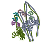
|
|---|---|
| 1 |
|
-要素
| #1: 抗体 | 分子量: 229148.250 Da / 分子数: 2 / 由来タイプ: 天然 由来: (天然) 参照: UniProt: G1N5L2*PLUS, myosin ATPase #2: タンパク質 | 分子量: 16989.145 Da / 分子数: 2 / 由来タイプ: 天然 由来: (天然) 参照: UniProt: Q6W5H0 #3: タンパク質 | 分子量: 19872.244 Da / 分子数: 2 / 由来タイプ: 天然 由来: (天然) 参照: UniProt: G3URE9 |
|---|
-実験情報
-実験
| 実験 | 手法: 電子顕微鏡法 |
|---|---|
| EM実験 | 試料の集合状態: PARTICLE / 3次元再構成法: 単粒子再構成法 |
- 試料調製
試料調製
| 構成要素 | 名称: Smooth muscle myosin II / タイプ: COMPLEX / 詳細: Whole myosin II isolated from turkey gizzard / Entity ID: all / 由来: NATURAL | ||||||||||||||||||||||||||||||
|---|---|---|---|---|---|---|---|---|---|---|---|---|---|---|---|---|---|---|---|---|---|---|---|---|---|---|---|---|---|---|---|
| 分子量 | 値: 0.53 MDa / 実験値: NO | ||||||||||||||||||||||||||||||
| 由来(天然) | 生物種: | ||||||||||||||||||||||||||||||
| 緩衝液 | pH: 7.5 | ||||||||||||||||||||||||||||||
| 緩衝液成分 |
| ||||||||||||||||||||||||||||||
| 試料 | 濃度: 0.1 mg/ml / 包埋: NO / シャドウイング: NO / 染色: NO / 凍結: YES 詳細: The specimen was cross-linked in presence of 0.1% glutaraldehyde for 60 seconds. | ||||||||||||||||||||||||||||||
| 試料支持 | グリッドの材料: COPPER / グリッドのサイズ: 300 divisions/in. / グリッドのタイプ: Quantifoil R1.2/1.3 | ||||||||||||||||||||||||||||||
| 急速凍結 | 装置: FEI VITROBOT MARK IV / 凍結剤: ETHANE / 湿度: 95 % / 凍結前の試料温度: 283.15 K |
- 電子顕微鏡撮影
| 実験機器 |  モデル: Titan Krios / 画像提供: FEI Company |
|---|---|
| 顕微鏡 | モデル: FEI TITAN KRIOS |
| 電子銃 | 電子線源:  FIELD EMISSION GUN / 加速電圧: 300 kV / 照射モード: FLOOD BEAM FIELD EMISSION GUN / 加速電圧: 300 kV / 照射モード: FLOOD BEAM |
| 電子レンズ | モード: BRIGHT FIELD / 倍率(公称値): 105000 X / 最大 デフォーカス(公称値): 3500 nm / 最小 デフォーカス(公称値): 1500 nm / Cs: 2.7 mm / C2レンズ絞り径: 70 µm / アライメント法: COMA FREE |
| 試料ホルダ | 凍結剤: NITROGEN 試料ホルダーモデル: FEI TITAN KRIOS AUTOGRID HOLDER |
| 撮影 | 電子線照射量: 43.3 e/Å2 / フィルム・検出器のモデル: GATAN K3 (6k x 4k) / 撮影したグリッド数: 2 / 実像数: 10951 |
| 画像スキャン | 横: 5760 / 縦: 4092 |
- 解析
解析
| ソフトウェア |
| |||||||||||||||||||||||||||||||||||||||||||||||||||||||
|---|---|---|---|---|---|---|---|---|---|---|---|---|---|---|---|---|---|---|---|---|---|---|---|---|---|---|---|---|---|---|---|---|---|---|---|---|---|---|---|---|---|---|---|---|---|---|---|---|---|---|---|---|---|---|---|---|
| EMソフトウェア |
| |||||||||||||||||||||||||||||||||||||||||||||||||||||||
| CTF補正 | タイプ: PHASE FLIPPING AND AMPLITUDE CORRECTION | |||||||||||||||||||||||||||||||||||||||||||||||||||||||
| 粒子像の選択 | 選択した粒子像数: 1765220 | |||||||||||||||||||||||||||||||||||||||||||||||||||||||
| 対称性 | 点対称性: C1 (非対称) | |||||||||||||||||||||||||||||||||||||||||||||||||||||||
| 3次元再構成 | 解像度: 4.3 Å / 解像度の算出法: FSC 0.143 CUT-OFF / 粒子像の数: 168613 / アルゴリズム: BACK PROJECTION / クラス平均像の数: 3 / 対称性のタイプ: POINT | |||||||||||||||||||||||||||||||||||||||||||||||||||||||
| 原子モデル構築 | プロトコル: RIGID BODY FIT / 空間: REAL | |||||||||||||||||||||||||||||||||||||||||||||||||||||||
| 原子モデル構築 |
| |||||||||||||||||||||||||||||||||||||||||||||||||||||||
| 精密化 | 交差検証法: NONE 立体化学のターゲット値: GeoStd + Monomer Library + CDL v1.2 | |||||||||||||||||||||||||||||||||||||||||||||||||||||||
| 原子変位パラメータ | Biso mean: 257.18 Å2 | |||||||||||||||||||||||||||||||||||||||||||||||||||||||
| 拘束条件 |
|