National Institutes of Health/National Institute of Arthritis and Musculoskeletal and Skin Diseases (NIH/NIAMS)
R01AR072036
United States
National Institutes of Health/National Institute of Arthritis and Musculoskeletal and Skin Diseases (NIH/NIAMS)
R01AR067279
United States
National Institutes of Health/National Heart, Lung, and Blood Institute (NIH/NHLBI)
R01HL139883
United States
Citation
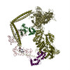
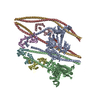
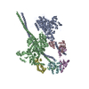
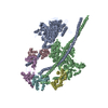
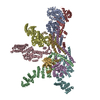
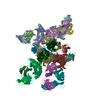
Journal: Nature / Year: 2020 Title: Cryo-EM structure of the inhibited (10S) form of myosin II. Authors: Shixin Yang / Prince Tiwari / Kyoung Hwan Lee / Osamu Sato / Mitsuo Ikebe / Raúl Padrón / Roger Craig / Abstract: Myosin II is the motor protein that enables muscle cells to contract and nonmuscle cells to move and change shape. The molecule has two identical heads attached to an elongated tail, and can exist in ...Myosin II is the motor protein that enables muscle cells to contract and nonmuscle cells to move and change shape. The molecule has two identical heads attached to an elongated tail, and can exist in two conformations: 10S and 6S, named for their sedimentation coefficients. The 6S conformation has an extended tail and assembles into polymeric filaments, which pull on actin filaments to generate force and motion. In 10S myosin, the tail is folded into three segments and the heads bend back and interact with each other and the tail, creating a compact conformation in which ATPase activity, actin activation and filament assembly are all highly inhibited. This switched-off structure appears to function as a key energy-conserving storage molecule in muscle and nonmuscle cells, which can be activated to form functional filaments as needed-but the mechanism of its inhibition is not understood. Here we have solved the structure of smooth muscle 10S myosin by cryo-electron microscopy with sufficient resolution to enable improved understanding of the function of the head and tail regions of the molecule and of the key intramolecular contacts that cause inhibition. Our results suggest an atomic model for the off state of myosin II, for its activation and unfolding by phosphorylation, and for understanding the clustering of disease-causing mutations near sites of intramolecular interaction.
Conc.: 0.1 mg/ml / Embedding applied: NO / Shadowing applied: NO / Staining applied: NO / Vitrification applied: YES Details: The specimen was cross-linked in presence of 0.1% glutaraldehyde for 60 seconds.
Electron dose: 43.3 e/Å2 / Film or detector model: GATAN K3 (6k x 4k) / Num. of grids imaged: 2 / Num. of real images: 10951
Image scans
Width: 5760 / Height: 4092
-
Processing
Software
Name
Version
Classification
phenix.real_space_refine
1.17.1_3660
refinement
PHENIX
1.17.1_3660
refinement
EM software
ID
Name
Version
Category
Details (eV)
1
RELION
3
particleselection
2
SerialEM
3.8.0
imageacquisition
4
CTFFIND
4.1.13
CTFcorrection
7
UCSF Chimera
1.14
modelfitting
Fitmodelinmap
9
RELION
3
initialEulerassignment
10
RELION
3
finalEulerassignment
11
RELION
3
classification
12
RELION
3
3Dreconstruction
19
Coot
0.8.9.2EL
modelrefinement
Cootwasusedforregularizationandlocalrefinement
20
PHENIX
1.17.1 - 3660
modelrefinement
ItwasusedforRealspacerefinementandmodelvalidation
CTF correction
Type: PHASE FLIPPING AND AMPLITUDE CORRECTION
Particle selection
Num. of particles selected: 1765220
Symmetry
Point symmetry: C1 (asymmetric)
3D reconstruction
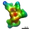
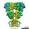
Resolution: 4.3 Å / Resolution method: FSC 0.143 CUT-OFF / Num. of particles: 168613 / Algorithm: BACK PROJECTION / Num. of class averages: 3 / Symmetry type: POINT
In the structure databanks used in Yorodumi, some data are registered as the other names, "COVID-19 virus" and "2019-nCoV". Here are the details of the virus and the list of structure data.
Jan 31, 2019. EMDB accession codes are about to change! (news from PDBe EMDB page)
EMDB accession codes are about to change! (news from PDBe EMDB page)
The allocation of 4 digits for EMDB accession codes will soon come to an end. Whilst these codes will remain in use, new EMDB accession codes will include an additional digit and will expand incrementally as the available range of codes is exhausted. The current 4-digit format prefixed with “EMD-” (i.e. EMD-XXXX) will advance to a 5-digit format (i.e. EMD-XXXXX), and so on. It is currently estimated that the 4-digit codes will be depleted around Spring 2019, at which point the 5-digit format will come into force.
The EM Navigator/Yorodumi systems omit the EMD- prefix.
Related info.:Q: What is EMD? / ID/Accession-code notation in Yorodumi/EM Navigator
Yorodumi is a browser for structure data from EMDB, PDB, SASBDB, etc.
This page is also the successor to EM Navigator detail page, and also detail information page/front-end page for Omokage search.
The word "yorodu" (or yorozu) is an old Japanese word meaning "ten thousand". "mi" (miru) is to see.
Related info.:EMDB / PDB / SASBDB / Comparison of 3 databanks / Yorodumi Search / Aug 31, 2016. New EM Navigator & Yorodumi / Yorodumi Papers / Jmol/JSmol / Function and homology information / Changes in new EM Navigator and Yorodumi
 Movie
Movie Controller


 Open data
 Basic information
 Components
 Keywords
 Function and homology information
 Authors
 United States, 3items
  Citation
 Structure visualization
 Downloads & links
 Other downloads
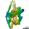

 PDBj
PDBj


 Assembly
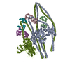
 Sample preparation
 Electron microscopy imaging
 FIELD EMISSION GUN / Accelerating voltage: 300 kV / Illumination mode: FLOOD BEAM
 Processing