 Movie
Movie Controller

[English] 日本語
 Yorodumi
- PDB-6wjv: Structure of the Saccharomyces cerevisiae polymerase epsilon holo... -
+ Open data

- Basic information
| Entry | Database: PDB / ID: 6wjv | ||||||||||||
|---|---|---|---|---|---|---|---|---|---|---|---|---|---|
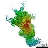
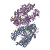
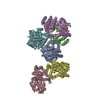
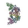
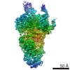
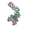
| Title | Structure of the Saccharomyces cerevisiae polymerase epsilon holoenzyme | ||||||||||||
 Components Components |
| ||||||||||||
 Keywords Keywords | CELL CYCLE / DNA replication / DNA polymerase | ||||||||||||
| Function / homology |  Function and homology information Function and homology informationregulation of DNA double-strand break processing / CHRAC / gene conversion / DNA-templated DNA replication maintenance of fidelity / DNA replication initiation / epsilon DNA polymerase complex / nucleotide-excision repair, DNA gap filling / SUMO binding / Activation of the pre-replicative complex / DNA replication proofreading ...regulation of DNA double-strand break processing / CHRAC / gene conversion / DNA-templated DNA replication maintenance of fidelity / DNA replication initiation / epsilon DNA polymerase complex / nucleotide-excision repair, DNA gap filling / SUMO binding / Activation of the pre-replicative complex / DNA replication proofreading / : / single-stranded DNA 3'-5' DNA exonuclease activity / mitotic DNA replication checkpoint signaling / mitotic intra-S DNA damage checkpoint signaling / mitotic sister chromatid cohesion / Hydrolases; Acting on ester bonds; Exodeoxyribonucleases producing 5'-phosphomonoesters / nucleosomal DNA binding / leading strand elongation / nuclear replication fork / Dual incision in TC-NER / error-prone translesion synthesis / base-excision repair, gap-filling / replication fork / chromatin DNA binding / base-excision repair / DNA-templated DNA replication / double-strand break repair via nonhomologous end joining / double-strand break repair / mitotic cell cycle / heterochromatin formation / single-stranded DNA binding / 4 iron, 4 sulfur cluster binding / double-stranded DNA binding / DNA-directed DNA polymerase / DNA-directed DNA polymerase activity / chromatin remodeling / protein heterodimerization activity / nucleotide binding / mRNA binding / DNA damage response / regulation of DNA-templated transcription / DNA binding / zinc ion binding / nucleus / cytoplasm Similarity search - Function | ||||||||||||
| Biological species |  | ||||||||||||
| Method | ELECTRON MICROSCOPY / single particle reconstruction / cryo EM / Resolution: 3.5 Å | ||||||||||||
 Authors Authors | Yuan, Z. / Georgescu, R. / Schauer, G.D. / O'Donnell, M. / Li, H. | ||||||||||||
| Funding support |  United States, 3items United States, 3items
| ||||||||||||
 Citation Citation | Journal: Nat Commun / Year: 2020 Title: Structure of the polymerase ε holoenzyme and atomic model of the leading strand replisome. Authors: Zuanning Yuan / Roxana Georgescu / Grant D Schauer / Michael E O'Donnell / Huilin Li / Abstract: The eukaryotic leading strand DNA polymerase (Pol) ε contains 4 subunits, Pol2, Dpb2, Dpb3 and Dpb4. Pol2 is a fusion of two B-family Pols; the N-terminal Pol module is catalytic and the C-terminal ...The eukaryotic leading strand DNA polymerase (Pol) ε contains 4 subunits, Pol2, Dpb2, Dpb3 and Dpb4. Pol2 is a fusion of two B-family Pols; the N-terminal Pol module is catalytic and the C-terminal Pol module is non-catalytic. Despite extensive efforts, there is no atomic structure for Pol ε holoenzyme, critical to understanding how DNA synthesis is coordinated with unwinding and the DNA path through the CMG helicase-Pol ε-PCNA clamp. We show here a 3.5-Å cryo-EM structure of yeast Pol ε revealing that the Dpb3-Dpb4 subunits bridge the two DNA Pol modules of Pol2, holding them rigid. This information enabled an atomic model of the leading strand replisome. Interestingly, the model suggests that an OB fold in Dbp2 directs leading ssDNA from CMG to the Pol ε active site. These results complete the DNA path from entry of parental DNA into CMG to exit of daughter DNA from PCNA. | ||||||||||||
| History |
|
- Structure visualization
| Movie |
Movie viewer |
|---|---|
| Structure viewer | Molecule: MolmilJmol/JSmol |
- Downloads & links
-Download
| PDBx/mmCIF format | 6wjv.cif.gz | 465.5 KB | Display | PDBx/mmCIF format |
|---|---|---|---|---|
| PDB format | pdb6wjv.ent.gz | 360.1 KB | Display | PDB format |
| PDBx/mmJSON format | 6wjv.json.gz | Tree view | PDBx/mmJSON format | |
| Others |  Other downloads Other downloads |
-Validation report
| Arichive directory | https://data.pdbj.org/pub/pdb/validation_reports/wj/6wjvftp://data.pdbj.org/pub/pdb/validation_reports/wj/6wjv | HTTPS FTP |
|---|
-Related structure data
| Related structure data |  21701MC M: map data used to model this data C: citing same article ( |
|---|---|
| Similar structure data |

-Links
 PDBj
PDBj


- Assembly
| Deposited unit | 
|
|---|---|
| 1 |
|
-Components
| #1: Protein | Mass: 255992.484 Da / Num. of mol.: 1 Source method: isolated from a genetically manipulated source Source: (gene. exp.) Strain: ATCC 204508 / S288c / Gene: POL2, DUN2, YNL262W, N0825 / Production host: References: UniProt: P21951, DNA-directed DNA polymerase, Hydrolases; Acting on ester bonds; Exodeoxyribonucleases producing 5'-phosphomonoesters |
|---|---|
| #2: Protein | Mass: 78425.852 Da / Num. of mol.: 1 Source method: isolated from a genetically manipulated source Source: (gene. exp.) Strain: ATCC 204508 / S288c / Gene: DPB2, YPR175W, P9705.7 / Production host: |
| #3: Protein | Mass: 22694.014 Da / Num. of mol.: 1 Source method: isolated from a genetically manipulated source Source: (gene. exp.) Strain: ATCC 204508 / S288c / Gene: DPB3, YBR278W, YBR2015 / Production host: |
| #4: Protein | Mass: 22029.484 Da / Num. of mol.: 1 Source method: isolated from a genetically manipulated source Source: (gene. exp.) Strain: ATCC 204508 / S288c / Gene: DPB4, YDR121W / Production host: |
| #5: Chemical | ChemComp-ZN /   Mass: 65.409 Da / Num. of mol.: 1 / Source method: obtained synthetically / Formula: Zn / Feature type: SUBJECT OF INVESTIGATION Mass: 65.409 Da / Num. of mol.: 1 / Source method: obtained synthetically / Formula: Zn / Feature type: SUBJECT OF INVESTIGATION |
| Has ligand of interest | Y |
| Has protein modification | Y |
-Experimental details
-Experiment
| Experiment | Method: ELECTRON MICROSCOPY |
|---|---|
| EM experiment | Aggregation state: PARTICLE / 3D reconstruction method: single particle reconstruction |
- Sample preparation
| Component | Name: DNA polymerase epsilon / Type: COMPLEX / Entity ID: #1-#4 / Source: RECOMBINANT |
|---|---|
| Source (natural) | Organism: Strain: ATCC 204508 / S288c |
| Source (recombinant) | Organism: |
| Buffer solution | pH: 7.5 |
| Specimen | Embedding applied: NO / Shadowing applied: NO / Staining applied: NO / Vitrification applied: YES |
| Vitrification | Cryogen name: ETHANE |
- Electron microscopy imaging
| Experimental equipment |  Model: Titan Krios / Image courtesy: FEI Company |
|---|---|
| Microscopy | Model: FEI TITAN KRIOS |
| Electron gun | Electron source:  FIELD EMISSION GUN / Accelerating voltage: 300 kV / Illumination mode: FLOOD BEAM FIELD EMISSION GUN / Accelerating voltage: 300 kV / Illumination mode: FLOOD BEAM |
| Electron lens | Mode: BRIGHT FIELD |
| Image recording | Electron dose: 60 e/Å2 / Detector mode: SUPER-RESOLUTION / Film or detector model: GATAN K2 SUMMIT (4k x 4k) |
- Processing
| CTF correction | Type: PHASE FLIPPING AND AMPLITUDE CORRECTION |
|---|---|
| Symmetry | Point symmetry: C1 (asymmetric) |
| 3D reconstruction | Resolution: 3.5 Å / Resolution method: FSC 0.143 CUT-OFF / Num. of particles: 187298 / Symmetry type: POINT |
| Atomic model building | Protocol: FLEXIBLE FIT / Space: REAL |
