Movie
Movie Controller


+ Open data

- Basic information
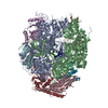
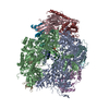
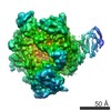
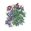
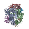
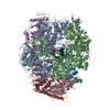
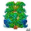
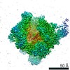
| Entry | Database: PDB / ID: 6vej | |||||||||||||||||||||
|---|---|---|---|---|---|---|---|---|---|---|---|---|---|---|---|---|---|---|---|---|---|---|
| Title | TriABC transporter from Pseudomonas aeruginosa | |||||||||||||||||||||
 Components Components |
| |||||||||||||||||||||
 Keywords Keywords | TRANSPORT PROTEIN / RND transporter / Pseudomonas aeruginosa / transmembrane / TriABC / TriC / PMF / cryoEM | |||||||||||||||||||||
| Function / homology |  Function and homology information Function and homology informationefflux pump complex / efflux transmembrane transporter activity / xenobiotic transmembrane transporter activity / transmembrane transporter activity / membrane => GO:0016020 / cell division / plasma membrane Similarity search - Function | |||||||||||||||||||||
| Biological species |   Pseudomonas aeruginosa (bacteria) Pseudomonas aeruginosa (bacteria) | |||||||||||||||||||||
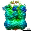
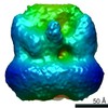
| Method | ELECTRON MICROSCOPY / single particle reconstruction / cryo EM / Resolution: 4.5 Å | |||||||||||||||||||||
 Authors Authors | Sygusch, J. / Fabre, L. / Rouiller, I. / Bhattacharyya, S. | |||||||||||||||||||||
| Funding support |  Canada, Canada,  United States, United States,  Australia, 6items Australia, 6items
| |||||||||||||||||||||
 Citation Citation | Journal: Structure / Year: 2021 Title: A "Drug Sweeping" State of the TriABC Triclosan Efflux Pump from Pseudomonas aeruginosa. Authors: Lucien Fabre / Abigail T Ntreh / Amira Yazidi / Inga V Leus / Jon W Weeks / Sudipta Bhattacharyya / Jakob Ruickoldt / Isabelle Rouiller / Helen I Zgurskaya / Jurgen Sygusch /   Abstract: The structure of the TriABC inner membrane component of the triclosan/SDS-specific efflux pump from Pseudomonas aeruginosa was determined by cryoelectron microscopy to 4.5 Å resolution. The ...The structure of the TriABC inner membrane component of the triclosan/SDS-specific efflux pump from Pseudomonas aeruginosa was determined by cryoelectron microscopy to 4.5 Å resolution. The complete structure of the inner membrane transporter TriC of the resistance-nodulation-division (RND) superfamily was solved, including a partial structure of the fused periplasmic membrane fusion subunits, TriA and TriB. The substrate-free conformation of TriABC represents an intermediate step in efflux complex assembly before the engagement of the outer membrane channel. Structural analysis identified a tunnel network whose constriction impedes substrate efflux, indicating inhibition of TriABC in the unengaged state. Blind docking studies revealed binding to TriC at the same loci by substrates and bulkier non-substrates. Together with functional analyses, we propose that selective substrate translocation involves conformational gating at the tunnel narrowing that, together with conformational ordering of TriA and TriB, creates an engaged state capable of mediating substrate efflux. | |||||||||||||||||||||
| History |
|
- Structure visualization
| Movie |
Movie viewer |
|---|---|
| Structure viewer | Molecule: MolmilJmol/JSmol |
- Downloads & links
-Download
| PDBx/mmCIF format | 6vej.cif.gz | 649.6 KB | Display | PDBx/mmCIF format |
|---|---|---|---|---|
| PDB format | pdb6vej.ent.gz | 538.5 KB | Display | PDB format |
| PDBx/mmJSON format | 6vej.json.gz | Tree view | PDBx/mmJSON format | |
| Others |  Other downloads Other downloads |
-Validation report
| Summary document | 6vej_validation.pdf.gz | 1.1 MB | Display | wwPDB validaton report |
|---|---|---|---|---|
| Full document | 6vej_full_validation.pdf.gz | 1.2 MB | Display | |
| Data in XML | 6vej_validation.xml.gz | 109.5 KB | Display | |
| Data in CIF | 6vej_validation.cif.gz | 157.4 KB | Display | |
| Arichive directory | https://data.pdbj.org/pub/pdb/validation_reports/ve/6vejftp://data.pdbj.org/pub/pdb/validation_reports/ve/6vej | HTTPS FTP |
-Related structure data
| Related structure data | 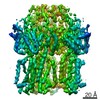 21363MC M: map data used to model this data C: citing same article ( |
|---|---|
| Similar structure data | |
| EM raw data | EMPIAR-10128 (Title: A "drug sweeping" state of the TriABC triclosan efflux pump from Pseudomonas aeruginosa Data size: 238.7 Data #1: Unaligned multi-frame micrographs [micrographs - multiframe] Data #2: Single particles stacks with polished particles [picked particles - multiframe - processed] Data #3: Relion repertory of the final refinement with a symmetry C3 [picked particles - single frame - processed] Data #4: Relion repertory of the final refinement with no symmetry [picked particles - multiframe - processed]) |
| Experimental dataset #1 | Data reference: 10.6019/EMPIAR-10128 / Data set type: EMPIAR |
-Links
 PDBj
PDBj


- Assembly
| Deposited unit | 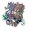
|
|---|---|
| 1 |
|
-Components
| #1: Protein | Mass: 112962.609 Da / Num. of mol.: 3 Source method: isolated from a genetically manipulated source Source: (gene. exp.) Pseudomonas aeruginosa (bacteria) / Gene: PA0158 / Production host: #2: Protein | Mass: 74070.781 Da / Num. of mol.: 3 Source method: isolated from a genetically manipulated source Source: (gene. exp.) Pseudomonas aeruginosa (bacteria) / Gene: PA0156, PA0157 / Production host: #3: Sugar | 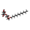  Type: D-saccharide / Mass: 510.615 Da / Num. of mol.: 3 / Source method: obtained synthetically / Formula: C24H46O11 / Comment: detergent*YM Type: D-saccharide / Mass: 510.615 Da / Num. of mol.: 3 / Source method: obtained synthetically / Formula: C24H46O11 / Comment: detergent*YM#4: Chemical | 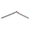  Mass: 594.992 Da / Num. of mol.: 3 / Source method: obtained synthetically / Formula: C38H74O4 Mass: 594.992 Da / Num. of mol.: 3 / Source method: obtained synthetically / Formula: C38H74O4#5: Chemical |   Mass: 256.424 Da / Num. of mol.: 3 / Source method: obtained synthetically / Formula: C16H32O2 Mass: 256.424 Da / Num. of mol.: 3 / Source method: obtained synthetically / Formula: C16H32O2Has ligand of interest | N | |
|---|
-Experimental details
-Experiment
| Experiment | Method: ELECTRON MICROSCOPY |
|---|---|
| EM experiment | Aggregation state: PARTICLE / 3D reconstruction method: single particle reconstruction |
- Sample preparation
| Component | Name: TriABC / Type: COMPLEX / Entity ID: #1-#2 / Source: RECOMBINANT |
|---|---|
| Molecular weight | Value: 0.560 MDa / Experimental value: YES |
| Source (natural) | Organism: Pseudomonas aeruginosa (bacteria) |
| Source (recombinant) | Organism: |
| Buffer solution | pH: 8 Details: 100 mM Tris HCl (pH 8.0), 150 mM NaCl, 1 mM PMSF, 0.03% (w/v) DDM. |
| Specimen | Embedding applied: NO / Shadowing applied: NO / Staining applied: NO / Vitrification applied: YES |
| Specimen support | Grid material: GRAPHENE OXIDE / Grid type: Quantifoil, UltrAuFoil, R1.2/1.3 |
| Vitrification | Cryogen name: ETHANE |
- Electron microscopy imaging
| Experimental equipment |  Model: Titan Krios / Image courtesy: FEI Company |
|---|---|
| Microscopy | Model: FEI TITAN KRIOS |
| Electron gun | Electron source:  FIELD EMISSION GUN / Accelerating voltage: 300 kV / Illumination mode: FLOOD BEAM FIELD EMISSION GUN / Accelerating voltage: 300 kV / Illumination mode: FLOOD BEAM |
| Electron lens | Mode: BRIGHT FIELD / Nominal magnification: 75000 X / Nominal defocus max: 3000 nm / Nominal defocus min: 1600 nm / Alignment procedure: COMA FREE |
| Specimen holder | Cryogen: NITROGEN / Specimen holder model: FEI TITAN KRIOS AUTOGRID HOLDER |
| Image recording | Electron dose: 50 e/Å2 / Film or detector model: FEI FALCON II (4k x 4k) / Num. of grids imaged: 1 / Num. of real images: 3483 Details: 1084 images kept for single particle analysis after exclusion of images that had no graphene oxide and proteins. |
- Processing
| EM software |
| |||||||||||||||||||||||||||||||||||
|---|---|---|---|---|---|---|---|---|---|---|---|---|---|---|---|---|---|---|---|---|---|---|---|---|---|---|---|---|---|---|---|---|---|---|---|---|
| CTF correction | Type: PHASE FLIPPING AND AMPLITUDE CORRECTION | |||||||||||||||||||||||||||||||||||
| Particle selection | Num. of particles selected: 35530 | |||||||||||||||||||||||||||||||||||
| 3D reconstruction | Resolution: 4.5 Å / Resolution method: FSC 0.143 CUT-OFF / Num. of particles: 35220 / Algorithm: BACK PROJECTION / Num. of class averages: 1 / Symmetry type: POINT | |||||||||||||||||||||||||||||||||||
| Atomic model building | Protocol: AB INITIO MODEL / Space: REAL Details: Subunits A-C were modeled by I-TASSER using CusA (3NE5) and then fitted into the EM density and refined with Phenix and model rebuilt using Coot. The N-terminal regions of subunits P-R were ...Details: Subunits A-C were modeled by I-TASSER using CusA (3NE5) and then fitted into the EM density and refined with Phenix and model rebuilt using Coot. The N-terminal regions of subunits P-R were modeled by I-TASSER based on MP domain of 3LNN while C-terminal regions of subunits P-R were modelled based on MP domain of 2V4D and similarly refined and model rebuilt. | |||||||||||||||||||||||||||||||||||
| Atomic model building |
|