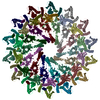
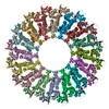
Cryo-EM reconstruction of Pyrobaculum filamentous virus 2 (PFV2)
Components
(A-DNA) x 2
Structural protein VP1
Structural protein VP2
Keywords
VIRUS / helical symmetry / archaeal pilus / STRUCTURAL PROTEIN
Function / homology
: / Pyrobaculum filamentous virus capsid protein / virion component / DNA binding / DNA / DNA (> 10) / DNA (> 100) / Major capsid protein 1 / Major capsid protein 2
Function and homology information
Biological species
Pyrobaculum filamentous virus 1
Method
ELECTRON MICROSCOPY / helical reconstruction / cryo EM / Resolution: 3.4 Å
National Institutes of Health/National Institute of General Medical Sciences (NIH/NIGMS)
R35GM122510
United States
Citation
Journal: Virus Evol / Year: 2020 Title: Structure of a filamentous virus uncovers familial ties within the archaeal virosphere. Authors: Fengbin Wang / Diana P Baquero / Zhangli Su / Tomasz Osinski / David Prangishvili / Edward H Egelman / Mart Krupovic / Abstract: Viruses infecting hyperthermophilic archaea represent one of the most enigmatic parts of the global virome, with viruses from different families showing no genomic relatedness to each other or to ...Viruses infecting hyperthermophilic archaea represent one of the most enigmatic parts of the global virome, with viruses from different families showing no genomic relatedness to each other or to viruses of bacteria and eukaryotes. Tristromaviruses, which build enveloped filamentous virions and infect hyperthermophilic neutrophiles of the order Thermoproteales, represent one such enigmatic virus families. They do not share genes with viruses from other families and have been believed to represent an evolutionarily independent virus lineage. A cryo-electron microscopic reconstruction of the tristromavirus Pyrobaculum filamentous virus 2 at 3.4 Å resolution shows that the virion is constructed from two paralogous major capsid proteins (MCP) which transform the linear dsDNA genome of the virus into A-form by tightly wrapping around it. Unexpectedly, the two MCP are homologous to the capsid proteins of other filamentous archaeal viruses, uncovering a deep evolutionary relationship within the archaeal virosphere.
1: A-DNA 2: A-DNA A: Structural protein VP1 B: Structural protein VP1 C: Structural protein VP1 D: Structural protein VP1 E: Structural protein VP1 F: Structural protein VP1 G: Structural protein VP1 H: Structural protein VP1 I: Structural protein VP1 J: Structural protein VP1 K: Structural protein VP1 L: Structural protein VP1 M: Structural protein VP1 N: Structural protein VP1 O: Structural protein VP1 P: Structural protein VP1 Q: Structural protein VP1 R: Structural protein VP1 S: Structural protein VP1 T: Structural protein VP1 U: Structural protein VP1 V: Structural protein VP1 W: Structural protein VP1 a: Structural protein VP2 b: Structural protein VP2 c: Structural protein VP2 d: Structural protein VP2 e: Structural protein VP2 f: Structural protein VP2 g: Structural protein VP2 h: Structural protein VP2 i: Structural protein VP2 j: Structural protein VP2 k: Structural protein VP2 l: Structural protein VP2 m: Structural protein VP2 n: Structural protein VP2 o: Structural protein VP2 p: Structural protein VP2 q: Structural protein VP2 r: Structural protein VP2 s: Structural protein VP2 t: Structural protein VP2 u: Structural protein VP2 v: Structural protein VP2 w: Structural protein VP2
Evidence: microscopy, helical filament was observed by negative staining and Cryo-EM
Type
Name
Symmetry operation
Number
identity operation
1_555
1
Buried area
316570 Å2
ΔGint
-1463 kcal/mol
Surface area
240570 Å2
Symmetry
Helical symmetry: (Circular symmetry: 1 / Dyad axis: no / N subunits divisor: 1 / Num. of operations: 23 / Rise per n subunits: 2.864 Å / Rotation per n subunits: 22.9482 °)
-
Components
#1: DNA chain
A-DNA
Mass: 99669.547 Da / Num. of mol.: 1 / Source method: isolated from a natural source / Source: (natural) Pyrobaculum filamentous virus 1
#2: DNA chain
A-DNA
Mass: 99660.539 Da / Num. of mol.: 1 / Source method: isolated from a natural source / Source: (natural) Pyrobaculum filamentous virus 1
#3: Protein
... StructuralproteinVP1
Mass: 14986.418 Da / Num. of mol.: 23 / Source method: isolated from a natural source / Source: (natural) Pyrobaculum filamentous virus 1 / References: UniProt: A0A140F3K6
#4: Protein
... StructuralproteinVP2
Mass: 15326.556 Da / Num. of mol.: 23 / Source method: isolated from a natural source / Source: (natural) Pyrobaculum filamentous virus 1 / References: UniProt: A0A140F3K7
Has protein modification
Y
-
Experimental details
-
Experiment
Experiment
Method: ELECTRON MICROSCOPY
EM experiment
Aggregation state: FILAMENT / 3D reconstruction method: helical reconstruction
In the structure databanks used in Yorodumi, some data are registered as the other names, "COVID-19 virus" and "2019-nCoV". Here are the details of the virus and the list of structure data.
Jan 31, 2019. EMDB accession codes are about to change! (news from PDBe EMDB page)
EMDB accession codes are about to change! (news from PDBe EMDB page)
The allocation of 4 digits for EMDB accession codes will soon come to an end. Whilst these codes will remain in use, new EMDB accession codes will include an additional digit and will expand incrementally as the available range of codes is exhausted. The current 4-digit format prefixed with “EMD-” (i.e. EMD-XXXX) will advance to a 5-digit format (i.e. EMD-XXXXX), and so on. It is currently estimated that the 4-digit codes will be depleted around Spring 2019, at which point the 5-digit format will come into force.
The EM Navigator/Yorodumi systems omit the EMD- prefix.
Related info.:Q: What is EMD? / ID/Accession-code notation in Yorodumi/EM Navigator
Yorodumi is a browser for structure data from EMDB, PDB, SASBDB, etc.
This page is also the successor to EM Navigator detail page, and also detail information page/front-end page for Omokage search.
The word "yorodu" (or yorozu) is an old Japanese word meaning "ten thousand". "mi" (miru) is to see.
Related info.:EMDB / PDB / SASBDB / Comparison of 3 databanks / Yorodumi Search / Aug 31, 2016. New EM Navigator & Yorodumi / Yorodumi Papers / Jmol/JSmol / Function and homology information / Changes in new EM Navigator and Yorodumi
 Movie
Movie Controller


 Open data
 Basic information
 Components
 Keywords
 Function and homology information
 Pyrobaculum filamentous virus 1
 Authors
 United States, 1items
  Citation
 Structure visualization
 Downloads & links
 Other downloads


 PDBj
PDBj


 Assembly
 Sample preparation
 Electron microscopy imaging
 FIELD EMISSION GUN / Accelerating voltage: 300 kV / Illumination mode: FLOOD BEAM
 Processing