 Movie
Movie Controller

[English] 日本語
 Yorodumi
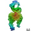
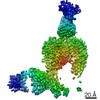
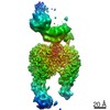
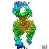
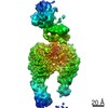
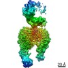
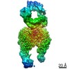
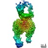
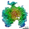
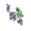
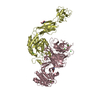
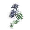
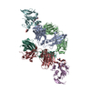
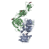
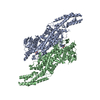
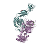
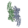
- PDB-6ujc: Integrin alpha-v beta-8 in complex with the Fabs C6-RGD3 and 11D12v2 -
+ Open data

- Basic information
| Entry | Database: PDB / ID: 6ujc | ||||||||||||||||||||||||
|---|---|---|---|---|---|---|---|---|---|---|---|---|---|---|---|---|---|---|---|---|---|---|---|---|---|
| Title | Integrin alpha-v beta-8 in complex with the Fabs C6-RGD3 and 11D12v2 | ||||||||||||||||||||||||
 Components Components |
| ||||||||||||||||||||||||
 Keywords Keywords | SIGNALING PROTEIN/IMMUNE SYSTEM / glycoprotein / adhesion / signaling / SIGNALING PROTEIN-IMMUNE SYSTEM complex | ||||||||||||||||||||||||
| Function / homology |  Function and homology information Function and homology informationganglioside metabolic process / Langerhans cell differentiation / integrin alphav-beta8 complex / integrin alphav-beta6 complex / hard palate development / transforming growth factor beta production / negative regulation of entry of bacterium into host cell / integrin alphav-beta5 complex / opsonin binding / integrin alphav-beta1 complex ...ganglioside metabolic process / Langerhans cell differentiation / integrin alphav-beta8 complex / integrin alphav-beta6 complex / hard palate development / transforming growth factor beta production / negative regulation of entry of bacterium into host cell / integrin alphav-beta5 complex / opsonin binding / integrin alphav-beta1 complex / Cross-presentation of particulate exogenous antigens (phagosomes) / extracellular matrix protein binding / Laminin interactions / placenta blood vessel development / negative regulation of lipoprotein metabolic process / integrin alphav-beta3 complex / negative regulation of low-density lipoprotein receptor activity / alphav-beta3 integrin-PKCalpha complex / alphav-beta3 integrin-HMGB1 complex / regulation of phagocytosis / negative regulation of lipid transport / Elastic fibre formation / alphav-beta3 integrin-IGF-1-IGF1R complex / transforming growth factor beta binding / entry into host cell by a symbiont-containing vacuole / positive regulation of small GTPase mediated signal transduction / filopodium membrane / extracellular matrix binding / apolipoprotein A-I-mediated signaling pathway / apoptotic cell clearance / wound healing, spreading of epidermal cells / cartilage development / positive regulation of intracellular signal transduction / heterotypic cell-cell adhesion / integrin complex / Molecules associated with elastic fibres / microvillus membrane / cell adhesion mediated by integrin / negative chemotaxis / Syndecan interactions / cell-substrate adhesion / endodermal cell differentiation / positive regulation of osteoblast proliferation / TGF-beta receptor signaling activates SMADs / PECAM1 interactions / lamellipodium membrane / fibronectin binding / negative regulation of macrophage derived foam cell differentiation / negative regulation of lipid storage / ECM proteoglycans / voltage-gated calcium channel activity / Integrin cell surface interactions / vasculogenesis / specific granule membrane / phagocytic vesicle / coreceptor activity / extrinsic apoptotic signaling pathway in absence of ligand / ERK1 and ERK2 cascade / positive regulation of cell adhesion / cell-matrix adhesion / substrate adhesion-dependent cell spreading / transforming growth factor beta receptor signaling pathway / Signal transduction by L1 / integrin-mediated signaling pathway / negative regulation of extrinsic apoptotic signaling pathway / calcium ion transmembrane transport / protein kinase C binding / response to virus / VEGFA-VEGFR2 Pathway / cell-cell adhesion / ruffle membrane / positive regulation of angiogenesis / cell migration / integrin binding / virus receptor activity / positive regulation of cytosolic calcium ion concentration / protease binding / angiogenesis / cell adhesion / positive regulation of cell migration / immune response / symbiont entry into host cell / external side of plasma membrane / negative regulation of gene expression / focal adhesion / positive regulation of cell population proliferation / Neutrophil degranulation / positive regulation of gene expression / cell surface / extracellular exosome / membrane / metal ion binding / plasma membrane / cytosol Similarity search - Function | ||||||||||||||||||||||||
| Biological species |  Homo sapiens (human) Homo sapiens (human) | ||||||||||||||||||||||||
| Method | ELECTRON MICROSCOPY / single particle reconstruction / cryo EM / Resolution: 3.56 Å | ||||||||||||||||||||||||
 Authors Authors | Campbell, M.G. / Cormier, A. / Cheng, Y. / Nishimura, S.L. | ||||||||||||||||||||||||
| Funding support |  United States, 7items United States, 7items
| ||||||||||||||||||||||||
 Citation Citation | Journal: Cell / Year: 2020 Title: Cryo-EM Reveals Integrin-Mediated TGF-β Activation without Release from Latent TGF-β. Authors: Melody G Campbell / Anthony Cormier / Saburo Ito / Robert I Seed / Andrew J Bondesson / Jianlong Lou / James D Marks / Jody L Baron / Yifan Cheng / Stephen L Nishimura / Abstract: Integrin αvβ8 binds with exquisite specificity to latent transforming growth factor-β (L-TGF-β). This binding is essential for activating L-TGF-β presented by a variety of cell types. ...Integrin αvβ8 binds with exquisite specificity to latent transforming growth factor-β (L-TGF-β). This binding is essential for activating L-TGF-β presented by a variety of cell types. Inhibiting αvβ8-mediated TGF-β activation blocks immunosuppressive regulatory T cell differentiation, which is a potential therapeutic strategy in cancer. Using cryo-electron microscopy, structure-guided mutagenesis, and cell-based assays, we reveal the binding interactions between the entire αvβ8 ectodomain and its intact natural ligand, L-TGF-β, as well as two different inhibitory antibody fragments to understand the structural underpinnings of αvβ8 binding specificity and TGF-β activation. Our studies reveal a mechanism of TGF-β activation where mature TGF-β signals within the confines of L-TGF-β and the release and diffusion of TGF-β are not required. The structural details of this mechanism provide a rational basis for therapeutic strategies to inhibit αvβ8-mediated L-TGF-β activation. | ||||||||||||||||||||||||
| History |
|
- Structure visualization
| Movie |
Movie viewer |
|---|---|
| Structure viewer | Molecule: MolmilJmol/JSmol |
- Downloads & links
-Download
| PDBx/mmCIF format | 6ujc.cif.gz | 265.4 KB | Display | PDBx/mmCIF format |
|---|---|---|---|---|
| PDB format | pdb6ujc.ent.gz | 194.5 KB | Display | PDB format |
| PDBx/mmJSON format | 6ujc.json.gz | Tree view | PDBx/mmJSON format | |
| Others |  Other downloads Other downloads |
-Validation report
| Summary document | 6ujc_validation.pdf.gz | 1 MB | Display | wwPDB validaton report |
|---|---|---|---|---|
| Full document | 6ujc_full_validation.pdf.gz | 1 MB | Display | |
| Data in XML | 6ujc_validation.xml.gz | 35.8 KB | Display | |
| Data in CIF | 6ujc_validation.cif.gz | 55 KB | Display | |
| Arichive directory | https://data.pdbj.org/pub/pdb/validation_reports/uj/6ujcftp://data.pdbj.org/pub/pdb/validation_reports/uj/6ujc | HTTPS FTP |
-Related structure data
| Related structure data | 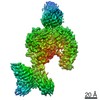 20796MC  6ujaC  6ujbC M: map data used to model this data C: citing same article ( |
|---|---|
| Similar structure data |
-Links
 PDBj
PDBj


- Assembly
| Deposited unit | 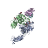
|
|---|---|
| 1 |
|
-Components
-Protein , 2 types, 2 molecules AB
| #1: Protein | Mass: 112813.352 Da / Num. of mol.: 1 Source method: isolated from a genetically manipulated source Source: (gene. exp.) Homo sapiens (human) / Gene: ITGAV, MSK8, VNRA, VTNR / Cell line (production host): CHO lec 3.2.8.1 / Production host:  Cricetulus griseus (Chinese hamster) / Tissue (production host): ovary / References: UniProt: P06756 Cricetulus griseus (Chinese hamster) / Tissue (production host): ovary / References: UniProt: P06756 |
|---|---|
| #2: Protein | Mass: 81276.664 Da / Num. of mol.: 1 Source method: isolated from a genetically manipulated source Source: (gene. exp.) Homo sapiens (human) / Gene: ITGB8 / Cell line (production host): CHO lec 3.2.8.1 / Production host: Cricetulus griseus (Chinese hamster) / Tissue (production host): ovary / References: UniProt: P26012 |
-Antibody , 2 types, 2 molecules EF
| #3: Antibody | Mass: 22785.475 Da / Num. of mol.: 1 Source method: isolated from a genetically manipulated source Source: (gene. exp.) Homo sapiens (human) |
|---|---|
| #4: Antibody | Mass: 24388.119 Da / Num. of mol.: 1 Source method: isolated from a genetically manipulated source Source: (gene. exp.) Homo sapiens (human) |
-Sugars , 3 types, 8 molecules 
| #5: Polysaccharide | alpha-D-mannopyranose-(1-2)-alpha-D-mannopyranose-(1-3)-[alpha-D-mannopyranose-(1-6)]beta-D- ...alpha-D-mannopyranose-(1-2)-alpha-D-mannopyranose-(1-3)-[alpha-D-mannopyranose-(1-6)]beta-D-mannopyranose-(1-4)-2-acetamido-2-deoxy-beta-D-glucopyranose-(1-4)-2-acetamido-2-deoxy-beta-D-glucopyranose Source method: isolated from a genetically manipulated source | ||
|---|---|---|---|
| #6: Polysaccharide | 2-acetamido-2-deoxy-beta-D-glucopyranose-(1-4)-2-acetamido-2-deoxy-beta-D-glucopyranose Source method: isolated from a genetically manipulated source #8: Sugar |  Type: D-saccharide, beta linking / Mass: 221.208 Da / Num. of mol.: 3 Type: D-saccharide, beta linking / Mass: 221.208 Da / Num. of mol.: 3Source method: isolated from a genetically manipulated source Formula: C8H15NO6 |
-Non-polymers , 2 types, 6 molecules 

| #7: Chemical | ChemComp-CA /  Mass: 40.078 Da / Num. of mol.: 5 / Source method: obtained synthetically / Formula: Ca Mass: 40.078 Da / Num. of mol.: 5 / Source method: obtained synthetically / Formula: Ca#9: Chemical | ChemComp-MG / |  Mass: 24.305 Da / Num. of mol.: 1 / Source method: obtained synthetically / Formula: Mg Mass: 24.305 Da / Num. of mol.: 1 / Source method: obtained synthetically / Formula: Mg |
|---|
-Details
| Has ligand of interest | N |
|---|---|
| Has protein modification | Y |
-Experimental details
-Experiment
| Experiment | Method: ELECTRON MICROSCOPY |
|---|---|
| EM experiment | Aggregation state: PARTICLE / 3D reconstruction method: single particle reconstruction |
- Sample preparation
| Component |
| ||||||||||||||||||||||||||||||
|---|---|---|---|---|---|---|---|---|---|---|---|---|---|---|---|---|---|---|---|---|---|---|---|---|---|---|---|---|---|---|---|
| Molecular weight | Value: 0.2 MDa / Experimental value: NO | ||||||||||||||||||||||||||||||
| Source (natural) |
| ||||||||||||||||||||||||||||||
| Source (recombinant) |
| ||||||||||||||||||||||||||||||
| Buffer solution | pH: 7.5 | ||||||||||||||||||||||||||||||
| Specimen | Embedding applied: NO / Shadowing applied: NO / Staining applied: NO / Vitrification applied: YES | ||||||||||||||||||||||||||||||
| Specimen support | Details: unspecified | ||||||||||||||||||||||||||||||
| Vitrification | Cryogen name: ETHANE |
- Electron microscopy imaging
| Experimental equipment |  Model: Titan Krios / Image courtesy: FEI Company |
|---|---|
| Microscopy | Model: FEI TITAN KRIOS |
| Electron gun | Electron source:  FIELD EMISSION GUN / Accelerating voltage: 300 kV / Illumination mode: FLOOD BEAM FIELD EMISSION GUN / Accelerating voltage: 300 kV / Illumination mode: FLOOD BEAM |
| Electron lens | Mode: BRIGHT FIELD |
| Image recording | Electron dose: 70 e/Å2 / Film or detector model: GATAN K2 SUMMIT (4k x 4k) |
- Processing
| CTF correction | Type: PHASE FLIPPING AND AMPLITUDE CORRECTION |
|---|---|
| Symmetry | Point symmetry: C1 (asymmetric) |
| 3D reconstruction | Resolution: 3.56 Å / Resolution method: FSC 0.143 CUT-OFF / Num. of particles: 221159 / Symmetry type: POINT |
