 Movie
Movie Controller


+ Open data

- Basic information
| Entry | Database: PDB / ID: 6tu3 | |||||||||
|---|---|---|---|---|---|---|---|---|---|---|
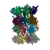
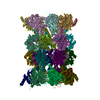
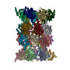
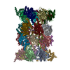
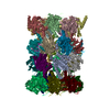
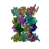
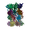
| Title | Rat 20S proteasome | |||||||||
 Components Components |
| |||||||||
 Keywords Keywords | HYDROLASE / PROTEASOME | |||||||||
| Function / homology |  Function and homology information Function and homology informationAntigen processing: Ub, ATP-independent proteasomal degradation / Cross-presentation of soluble exogenous antigens (endosomes) / Proteasome assembly / Regulation of ornithine decarboxylase (ODC) / Degradation of CRY and PER proteins / Oxygen-dependent proline hydroxylation of Hypoxia-inducible Factor Alpha / Autodegradation of Cdh1 by Cdh1:APC/C / SCF-beta-TrCP mediated degradation of Emi1 / APC/C:Cdc20 mediated degradation of Securin / APC/C:Cdh1 mediated degradation of Cdc20 and other APC/C:Cdh1 targeted proteins in late mitosis/early G1 ...Antigen processing: Ub, ATP-independent proteasomal degradation / Cross-presentation of soluble exogenous antigens (endosomes) / Proteasome assembly / Regulation of ornithine decarboxylase (ODC) / Degradation of CRY and PER proteins / Oxygen-dependent proline hydroxylation of Hypoxia-inducible Factor Alpha / Autodegradation of Cdh1 by Cdh1:APC/C / SCF-beta-TrCP mediated degradation of Emi1 / APC/C:Cdc20 mediated degradation of Securin / APC/C:Cdh1 mediated degradation of Cdc20 and other APC/C:Cdh1 targeted proteins in late mitosis/early G1 / Cdc20:Phospho-APC/C mediated degradation of Cyclin A / SCF(Skp2)-mediated degradation of p27/p21 / Autodegradation of the E3 ubiquitin ligase COP1 / Asymmetric localization of PCP proteins / Degradation of DVL / Hedgehog ligand biogenesis / Dectin-1 mediated noncanonical NF-kB signaling / Degradation of GLI1 by the proteasome / Hedgehog 'on' state / TNFR2 non-canonical NF-kB pathway / NIK-->noncanonical NF-kB signaling / Assembly of the pre-replicative complex / CDK-mediated phosphorylation and removal of Cdc6 / G2/M Checkpoints / Ubiquitin-Mediated Degradation of Phosphorylated Cdc25A / Ubiquitin-dependent degradation of Cyclin D / The role of GTSE1 in G2/M progression after G2 checkpoint / FBXL7 down-regulates AURKA during mitotic entry and in early mitosis / RUNX1 regulates transcription of genes involved in differentiation of HSCs / Regulation of RUNX3 expression and activity / GSK3B and BTRC:CUL1-mediated-degradation of NFE2L2 / GLI3 is processed to GLI3R by the proteasome / Activation of NF-kappaB in B cells / Degradation of beta-catenin by the destruction complex / Degradation of AXIN / UCH proteinases / Degradation of CDH1 / Regulation of RAS by GAPs / Orc1 removal from chromatin / Neddylation / AUF1 (hnRNP D0) binds and destabilizes mRNA / Regulation of PTEN stability and activity / KEAP1-NFE2L2 pathway / Separation of Sister Chromatids / MAPK6/MAPK4 signaling / ER-Phagosome pathway / Antigen processing: Ubiquitination & Proteasome degradation / ABC-family protein mediated transport / Ub-specific processing proteases / Neutrophil degranulation / proteasome core complex / myofibril / immune system process / proteasome endopeptidase complex / NF-kappaB binding / proteasome core complex, beta-subunit complex / threonine-type endopeptidase activity / proteasome core complex, alpha-subunit complex / : / proteasome complex / sarcomere / negative regulation of inflammatory response to antigenic stimulus / P-body / lipopolysaccharide binding / response to virus / nuclear matrix / peptidase activity / regulation of inflammatory response / response to oxidative stress / endopeptidase activity / proteasome-mediated ubiquitin-dependent protein catabolic process / positive regulation of canonical NF-kappaB signal transduction / cilium / nuclear body / ciliary basal body / ribosome / ubiquitin protein ligase binding / centrosome / mitochondrion / proteolysis / RNA binding / nucleoplasm / identical protein binding / nucleus / cytoplasm / cytosol Similarity search - Function | |||||||||
| Biological species |  | |||||||||
| Method | ELECTRON MICROSCOPY / single particle reconstruction / cryo EM / Resolution: 2.7 Å | |||||||||
 Authors Authors | Deshmukh, F.K. / Polkinghorn, C.R. / Elad, N. / Sharon, M. | |||||||||
| Funding support |  Israel, 2items Israel, 2items
| |||||||||
 Citation Citation | Journal: ACS Cent Sci / Year: 2020 Title: Comparative Structural Analysis of 20S Proteasome Ortholog Protein Complexes by Native Mass Spectrometry. Authors: Shay Vimer / Gili Ben-Nissan / David Morgenstern / Fanindra Kumar-Deshmukh / Caley Polkinghorn / Royston S Quintyn / Yury V Vasil'ev / Joseph S Beckman / Nadav Elad / Vicki H Wysocki / Michal Sharon /  Abstract: Ortholog protein complexes are responsible for equivalent functions in different organisms. However, during evolution, each organism adapts to meet its physiological needs and the environmental ...Ortholog protein complexes are responsible for equivalent functions in different organisms. However, during evolution, each organism adapts to meet its physiological needs and the environmental challenges imposed by its niche. This selection pressure leads to structural diversity in protein complexes, which are often difficult to specify, especially in the absence of high-resolution structures. Here, we describe a multilevel experimental approach based on native mass spectrometry (MS) tools for elucidating the structural preservation and variations among highly related protein complexes. The 20S proteasome, an essential protein degradation machinery, served as our model system, wherein we examined five complexes isolated from different organisms. We show that throughout evolution, from the archaeal prokaryotic complex to the eukaryotic 20S proteasomes in yeast () and mammals (rat - , rabbit - and human - HEK293 cells), the proteasome increased both in size and stability. Native MS structural signatures of the rat and rabbit 20S proteasomes, which heretofore lacked high-resolution, three-dimensional structures, highly resembled that of the human complex. Using cryoelectron microscopy single-particle analysis, we were able to obtain a high-resolution structure of the rat 20S proteasome, allowing us to validate the MS-based results. Our study also revealed that the yeast complex, and not those in mammals, was the largest in size and displayed the greatest degree of kinetic stability. Moreover, we also identified a new proteoform of the PSMA7 subunit that resides within the rat and rabbit complexes, which to our knowledge have not been previously described. Altogether, our strategy enables elucidation of the unique structural properties of protein complexes that are highly similar to one another, a framework that is valid not only to ortholog protein complexes, but also for other highly related protein assemblies. | |||||||||
| History |
|
- Structure visualization
| Movie |
Movie viewer |
|---|---|
| Structure viewer | Molecule: MolmilJmol/JSmol |
- Downloads & links
-Download
| PDBx/mmCIF format | 6tu3.cif.gz | 1 MB | Display | PDBx/mmCIF format |
|---|---|---|---|---|
| PDB format | pdb6tu3.ent.gz | 847.7 KB | Display | PDB format |
| PDBx/mmJSON format | 6tu3.json.gz | Tree view | PDBx/mmJSON format | |
| Others |  Other downloads Other downloads |
-Validation report
| Arichive directory | https://data.pdbj.org/pub/pdb/validation_reports/tu/6tu3ftp://data.pdbj.org/pub/pdb/validation_reports/tu/6tu3 | HTTPS FTP |
|---|
-Related structure data
| Related structure data | 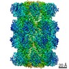 10586MC M: map data used to model this data C: citing same article ( |
|---|---|
| Similar structure data |

-Links
 PDBj
PDBj


- Assembly
| Deposited unit | 
|
|---|---|
| 1 |
|
-Components
-Proteasome subunit alpha type- ... , 7 types, 14 molecules AOBPCQDRESFTGU
| #1: Protein | Mass: 27432.459 Da / Num. of mol.: 2 / Source method: isolated from a natural source / Source: (natural) References: UniProt: P60901, proteasome endopeptidase complex #2: Protein | Mass: 25955.549 Da / Num. of mol.: 2 / Source method: isolated from a natural source / Source: (natural) References: UniProt: P17220, proteasome endopeptidase complex #3: Protein | Mass: 29539.830 Da / Num. of mol.: 2 / Source method: isolated from a natural source / Source: (natural) References: UniProt: P21670, proteasome endopeptidase complex #4: Protein | Mass: 28369.439 Da / Num. of mol.: 2 / Source method: isolated from a natural source / Source: (natural) References: UniProt: P48004, proteasome endopeptidase complex #5: Protein | Mass: 26416.850 Da / Num. of mol.: 2 / Source method: isolated from a natural source / Source: (natural) References: UniProt: P34064, proteasome endopeptidase complex #6: Protein | Mass: 29557.541 Da / Num. of mol.: 2 / Source method: isolated from a natural source / Source: (natural) References: UniProt: P18420, proteasome endopeptidase complex #7: Protein | Mass: 28456.275 Da / Num. of mol.: 2 / Source method: isolated from a natural source / Source: (natural) References: UniProt: P18422, proteasome endopeptidase complex |
|---|
-Proteasome subunit beta type- ... , 7 types, 14 molecules HVIWJXKYLZMaNb
| #8: Protein | Mass: 25309.576 Da / Num. of mol.: 2 / Source method: isolated from a natural source / Source: (natural) References: UniProt: P28073, proteasome endopeptidase complex #9: Protein | Mass: 29963.461 Da / Num. of mol.: 2 / Source method: isolated from a natural source / Source: (natural) References: UniProt: Q9JHW0, proteasome endopeptidase complex #10: Protein | Mass: 22988.895 Da / Num. of mol.: 2 / Source method: isolated from a natural source / Source: (natural) References: UniProt: P40112, proteasome endopeptidase complex #11: Protein | Mass: 22941.381 Da / Num. of mol.: 2 / Source method: isolated from a natural source / Source: (natural) References: UniProt: P40307, proteasome endopeptidase complex #12: Protein | Mass: 28615.408 Da / Num. of mol.: 2 / Source method: isolated from a natural source / Source: (natural) References: UniProt: P28075, proteasome endopeptidase complex #13: Protein | Mass: 26511.301 Da / Num. of mol.: 2 / Source method: isolated from a natural source / Source: (natural) References: UniProt: P18421, proteasome endopeptidase complex #14: Protein | Mass: 29226.248 Da / Num. of mol.: 2 / Source method: isolated from a natural source / Source: (natural) References: UniProt: P34067, proteasome endopeptidase complex |
|---|
-Experimental details
-Experiment
| Experiment | Method: ELECTRON MICROSCOPY |
|---|---|
| EM experiment | Aggregation state: PARTICLE / 3D reconstruction method: single particle reconstruction |
- Sample preparation
| Component | Name: Rat liver 20S proteasome / Type: COMPLEX Details: Endogenous 20S proteasome purified from the rat livers by anion-exchange chromatography Entity ID: all / Source: NATURAL |
|---|---|
| Source (natural) | Organism: |
| Buffer solution | pH: 7.4 |
| Buffer component | Conc.: 160 mM / Name: Potassium phosphate / Formula: K3PO4 |
| Specimen | Conc.: 14 mg/ml / Embedding applied: NO / Shadowing applied: NO / Staining applied: NO / Vitrification applied: YES / Details: Purified endogenous rat liver 20S proteasome |
| Specimen support | Details: Grids were glow discharged 30 minutes before applying the sample. Grid material: COPPER / Grid mesh size: 300 divisions/in. / Grid type: C-flat-2/2 |
| Vitrification | Instrument: FEI VITROBOT MARK IV / Cryogen name: ETHANE / Humidity: 100 % / Chamber temperature: 277 K / Details: blot for 3 seconds |
- Electron microscopy imaging
| Experimental equipment |  Model: Titan Krios / Image courtesy: FEI Company |
|---|---|
| Microscopy | Model: FEI TITAN KRIOS Details: Preliminary grid screening was performed on Talos Arctica |
| Electron gun | Electron source:  FIELD EMISSION GUN / Accelerating voltage: 300 kV / Illumination mode: OTHER FIELD EMISSION GUN / Accelerating voltage: 300 kV / Illumination mode: OTHER |
| Electron lens | Mode: DIFFRACTION / Nominal magnification: 105000 X / Nominal defocus max: 1600 nm / Nominal defocus min: 600 nm / Cs: 2.7 mm / C2 aperture diameter: 70 µm |
| Specimen holder | Cryogen: NITROGEN / Specimen holder model: FEI TITAN KRIOS AUTOGRID HOLDER |
| Image recording | Average exposure time: 1.5 sec. / Electron dose: 37 e/Å2 / Film or detector model: GATAN K3 BIOQUANTUM (6k x 4k) / Num. of grids imaged: 1 / Num. of real images: 2234 Details: Images were collected in movie-mode at 45 frames per second |
| EM imaging optics | Energyfilter name: GIF Bioquantum / Energyfilter slit width: 20 eV |
- Processing
| EM software |
| ||||||||||||||||||||||||||||
|---|---|---|---|---|---|---|---|---|---|---|---|---|---|---|---|---|---|---|---|---|---|---|---|---|---|---|---|---|---|
| CTF correction | Type: PHASE FLIPPING AND AMPLITUDE CORRECTION | ||||||||||||||||||||||||||||
| Particle selection | Num. of particles selected: 284161 | ||||||||||||||||||||||||||||
| Symmetry | Point symmetry: C2 (2 fold cyclic) | ||||||||||||||||||||||||||||
| 3D reconstruction | Resolution: 2.7 Å / Resolution method: FSC 0.143 CUT-OFF / Num. of particles: 245800 / Algorithm: BACK PROJECTION / Symmetry type: POINT | ||||||||||||||||||||||||||||
| Atomic model building | Protocol: FLEXIBLE FIT |
