 Movie
Movie Controller


+ Open data

- Basic information
| Entry | Database: PDB / ID: 6r4r | |||||||||||||||
|---|---|---|---|---|---|---|---|---|---|---|---|---|---|---|---|---|
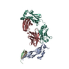
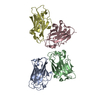
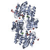
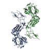
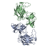
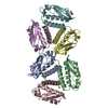
| Title | Cryo-EM Structure of the PI3-Kinase SH3 Domain Amyloid Fibril | |||||||||||||||
 Components Components | Phosphatidylinositol 3-kinase regulatory subunit alpha | |||||||||||||||
 Keywords Keywords | PROTEIN FIBRIL / amyloid fibril / sh3 domain | |||||||||||||||
| Function / homology |  Function and homology information Function and homology informationRHOC GTPase cycle / PI3K events in ERBB4 signaling / Interleukin-7 signaling / GAB1 signalosome / PI3K events in ERBB2 signaling / MET activates PI3K/AKT signaling / CDC42 GTPase cycle / : / RHOJ GTPase cycle / RAC3 GTPase cycle ...RHOC GTPase cycle / PI3K events in ERBB4 signaling / Interleukin-7 signaling / GAB1 signalosome / PI3K events in ERBB2 signaling / MET activates PI3K/AKT signaling / CDC42 GTPase cycle / : / RHOJ GTPase cycle / RAC3 GTPase cycle / Erythropoietin activates Phosphoinositide-3-kinase (PI3K) / FLT3 Signaling / RND2 GTPase cycle / RND1 GTPase cycle / IRS-mediated signalling / GPVI-mediated activation cascade / Signaling by SCF-KIT / Downstream signal transduction / PI3K/AKT activation / Signaling by ALK / Role of phospholipids in phagocytosis / Tie2 Signaling / Role of LAT2/NTAL/LAB on calcium mobilization / CD28 dependent PI3K/Akt signaling / : / RAC2 GTPase cycle / Interleukin receptor SHC signaling / RND3 GTPase cycle / Co-stimulation by ICOS / PI-3K cascade:FGFR1 / PI-3K cascade:FGFR2 / PI-3K cascade:FGFR3 / PI-3K cascade:FGFR4 / Antigen activates B Cell Receptor (BCR) leading to generation of second messengers / PI3K Cascade / PIP3 activates AKT signaling / GP1b-IX-V activation signalling / RAF/MAP kinase cascade / PI5P, PP2A and IER3 Regulate PI3K/AKT Signaling / Synthesis of PIPs at the plasma membrane / RHOA GTPase cycle / RHOF GTPase cycle / DAP12 signaling / RHOU GTPase cycle / RHOV GTPase cycle / Regulation of signaling by CBL / Downstream TCR signaling / RHOG GTPase cycle / RET signaling / Interleukin-3, Interleukin-5 and GM-CSF signaling / VEGFA-VEGFR2 Pathway / phosphatidylinositol 3-kinase regulator activity / 1-phosphatidylinositol-3-kinase regulator activity / positive regulation of endoplasmic reticulum unfolded protein response / phosphatidylinositol 3-kinase activator activity / ErbB-3 class receptor binding / transmembrane receptor protein tyrosine kinase adaptor activity / phosphatidylinositol 3-kinase complex / phosphatidylinositol 3-kinase complex, class IA / Extra-nuclear estrogen signaling / G alpha (q) signalling events / phosphatidylinositol phosphate biosynthetic process / insulin receptor substrate binding / enzyme-substrate adaptor activity / phosphatidylinositol 3-kinase binding / insulin-like growth factor receptor binding / substrate adhesion-dependent cell spreading / insulin-like growth factor receptor signaling pathway / intracellular glucose homeostasis / response to endoplasmic reticulum stress / positive regulation of RNA splicing / GTPase activator activity / positive regulation of D-glucose import across plasma membrane / insulin receptor binding / positive regulation of protein localization to plasma membrane / phosphatidylinositol 3-kinase/protein kinase B signal transduction / positive regulation of protein import into nucleus / cellular response to insulin stimulus / insulin receptor signaling pathway / protein transport / protein stabilization / negative regulation of apoptotic process / positive regulation of transcription by RNA polymerase II / identical protein binding / nucleus Similarity search - Function | |||||||||||||||
| Biological species |  | |||||||||||||||
| Method | ELECTRON MICROSCOPY / helical reconstruction / cryo EM / Resolution: 3.4 Å | |||||||||||||||
 Authors Authors | Roeder, C. / Vettore, N. / Mangels, L.N. / Gremer, L. / Ravelli, R.B.G. / Willbold, D. / Hoyer, W. / Buell, A.K. / Schroder, G.F. | |||||||||||||||
| Funding support |  Germany, 4items Germany, 4items
| |||||||||||||||
 Citation Citation | Journal: Nat Commun / Year: 2019 Title: Atomic structure of PI3-kinase SH3 amyloid fibrils by cryo-electron microscopy. Authors: Christine Röder / Nicola Vettore / Lena N Mangels / Lothar Gremer / Raimond B G Ravelli / Dieter Willbold / Wolfgang Hoyer / Alexander K Buell / Gunnar F Schröder /   Abstract: High resolution structural information on amyloid fibrils is crucial for the understanding of their formation mechanisms and for the rational design of amyloid inhibitors in the context of protein ...High resolution structural information on amyloid fibrils is crucial for the understanding of their formation mechanisms and for the rational design of amyloid inhibitors in the context of protein misfolding diseases. The Src-homology 3 domain of phosphatidyl-inositol-3-kinase (PI3K-SH3) is a model amyloid system that plays a pivotal role in our basic understanding of protein misfolding and aggregation. Here, we present the atomic model of the PI3K-SH3 amyloid fibril with a resolution determined to 3.4 Å by cryo-electron microscopy (cryo-EM). The fibril is composed of two intertwined protofilaments that create an interface spanning 13 residues from each monomer. The model comprises residues 1-77 out of 86 amino acids in total, with the missing residues located in the highly flexible C-terminus. The fibril structure allows us to rationalise the effects of chemically conservative point mutations as well as of the previously reported sequence perturbations on PI3K-SH3 fibril formation and growth. | |||||||||||||||
| History |
|
- Structure visualization
| Movie |
Movie viewer |
|---|---|
| Structure viewer | Molecule: MolmilJmol/JSmol |
- Downloads & links
-Download
| PDBx/mmCIF format | 6r4r.cif.gz | 103 KB | Display | PDBx/mmCIF format |
|---|---|---|---|---|
| PDB format | pdb6r4r.ent.gz | 77 KB | Display | PDB format |
| PDBx/mmJSON format | 6r4r.json.gz | Tree view | PDBx/mmJSON format | |
| Others |  Other downloads Other downloads |
-Validation report
| Arichive directory | https://data.pdbj.org/pub/pdb/validation_reports/r4/6r4rftp://data.pdbj.org/pub/pdb/validation_reports/r4/6r4r | HTTPS FTP |
|---|
-Related structure data
| Related structure data |  4727MC M: map data used to model this data C: citing same article ( |
|---|---|
| Similar structure data | |
| EM raw data | EMPIAR-11092 (Title: Micrographs of PI3-kinase SH3 amyloid fibrils / Data size: 32.0 Data #1: PI3-kinase SH3 amyloid fibrils, MotionCor2-aligned dose-weighted averages [micrographs - single frame] Data #2: PI3-kinase SH3 amyloid fibrils, MotionCor2-aligned non-dose-weighted averages [micrographs - single frame]) |


-Links
 PDBj
PDBj


- Assembly
| Deposited unit | 
|
|---|---|
| 1 |
|
-Components
| #1: Protein | Mass: 9649.585 Da / Num. of mol.: 7 Source method: isolated from a genetically manipulated source Source: (gene. exp.)  |
|---|
-Experimental details
-Experiment
| Experiment | Method: ELECTRON MICROSCOPY |
|---|---|
| EM experiment | Aggregation state: FILAMENT / 3D reconstruction method: helical reconstruction |
- Sample preparation
| Component | Name: Fibril of PI3K-SH3 domains / Type: COMPLEX / Entity ID: all / Source: RECOMBINANT |
|---|---|
| Molecular weight | Value: 34.55 kDa/nm / Experimental value: NO |
| Source (natural) | Organism: |
| Source (recombinant) | Organism: |
| Buffer solution | pH: 2.5 |
| Buffer component | Conc.: 0.11 mg/mL / Name: glycin-hydrochloride / Formula: C2H6ClNO2 |
| Specimen | Conc.: 1.62 mg/ml / Embedding applied: NO / Shadowing applied: NO / Staining applied: NO / Vitrification applied: YES |
| Specimen support | Grid material: COPPER / Grid mesh size: 300 divisions/in. / Grid type: Quantifoil R1.2/1.3 |
| Vitrification | Cryogen name: ETHANE |
- Electron microscopy imaging
| Experimental equipment |  Model: Talos Arctica / Image courtesy: FEI Company |
|---|---|
| Microscopy | Model: FEI TECNAI ARCTICA |
| Electron gun | Electron source:  FIELD EMISSION GUN / Accelerating voltage: 200 kV / Illumination mode: FLOOD BEAM FIELD EMISSION GUN / Accelerating voltage: 200 kV / Illumination mode: FLOOD BEAM |
| Electron lens | Mode: BRIGHT FIELD / Cs: 2.7 mm |
| Image recording | Average exposure time: 65 sec. / Electron dose: 26 e/Å2 / Detector mode: COUNTING / Film or detector model: FEI FALCON III (4k x 4k) / Num. of real images: 622 |
| Image scans | Width: 4096 / Height: 4096 |
- Processing
| EM software |
| ||||||||||||||||||||||||||||||||
|---|---|---|---|---|---|---|---|---|---|---|---|---|---|---|---|---|---|---|---|---|---|---|---|---|---|---|---|---|---|---|---|---|---|
| CTF correction | Type: PHASE FLIPPING AND AMPLITUDE CORRECTION | ||||||||||||||||||||||||||||||||
| Helical symmerty | Angular rotation/subunit: 179.436 ° / Axial rise/subunit: 2.3548 Å / Axial symmetry: C1 | ||||||||||||||||||||||||||||||||
| Particle selection | Num. of particles selected: 103733 / Details: Filaments were picked manually in Relion2 | ||||||||||||||||||||||||||||||||
| 3D reconstruction | Resolution: 3.4 Å / Resolution method: FSC 0.143 CUT-OFF / Num. of particles: 27681 Details: For the even/odd test, the segment images were split by entire fibrils and refined separately for 25 iterations using the same reference density (low-passed filtered to 20 A). Symmetry type: HELICAL | ||||||||||||||||||||||||||||||||
| Atomic model building | Protocol: AB INITIO MODEL / Space: REAL |
