 ムービー
ムービー コントローラー


+ データを開く

- 基本情報
基本情報
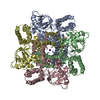
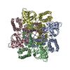
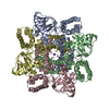
| 登録情報 | データベース: PDB / ID: 6qd0 | ||||||
|---|---|---|---|---|---|---|---|
| タイトル | MloK1 model from single particle analysis of 2D crystals, class 4 (compact/open conformation) | ||||||
 要素 要素 | Cyclic nucleotide-gated potassium channel mll3241 | ||||||
 キーワード キーワード | MEMBRANE PROTEIN / Voltage-gated potassium channel / Cyclic nucleotide-binding domain / ion channel / ion transport | ||||||
| 機能・相同性 |  機能・相同性情報 機能・相同性情報intracellularly cyclic nucleotide-activated monoatomic cation channel activity / potassium channel activity / cAMP binding / protein-containing complex binding / identical protein binding / plasma membrane 類似検索 - 分子機能 | ||||||
| 生物種 |  Mesorhizobium loti MAFF303099 (根粒菌) Mesorhizobium loti MAFF303099 (根粒菌) | ||||||
| 手法 | 電子顕微鏡法 / 単粒子再構成法 / クライオ電子顕微鏡法 / 解像度: 4.5 Å | ||||||
 データ登録者 データ登録者 | Righetto, R. / Biyani, N. / Kowal, J. / Chami, M. / Stahlberg, H. | ||||||
| 資金援助 |  スイス, 1件 スイス, 1件
| ||||||
 引用 引用 | ジャーナル: Nat Commun / 年: 2019 タイトル: Retrieving high-resolution information from disordered 2D crystals by single-particle cryo-EM. 著者: Ricardo D Righetto / Nikhil Biyani / Julia Kowal / Mohamed Chami / Henning Stahlberg / 要旨: Electron crystallography can reveal the structure of membrane proteins within 2D crystals under close-to-native conditions. High-resolution structural information can only be reached if crystals are ...Electron crystallography can reveal the structure of membrane proteins within 2D crystals under close-to-native conditions. High-resolution structural information can only be reached if crystals are perfectly flat and highly ordered. In practice, such crystals are difficult to obtain. Available image unbending algorithms correct for disorder, but only perform well on images of non-tilted, flat crystals, while out-of-plane distortions are not addressed. Here, we present an approach that employs single-particle refinement procedures to locally unbend crystals in 3D. With this method, density maps of the MloK1 potassium channel with a resolution of 4 Å were obtained from images of 2D crystals that do not diffract beyond 10 Å. Furthermore, 3D classification allowed multiple structures to be resolved, revealing a series of MloK1 conformations within a single 2D crystal. This conformational heterogeneity explains the poor diffraction observed and is related to channel function. The approach is implemented in the FOCUS package. | ||||||
| 履歴 |
|
- 構造の表示
構造の表示
| ムービー |
ムービービューア |
|---|---|
| 構造ビューア | 分子: MolmilJmol/JSmol |
- ダウンロードとリンク
-ダウンロード
| PDBx/mmCIF形式 | 6qd0.cif.gz | 445.7 KB | 表示 | PDBx/mmCIF形式 |
|---|---|---|---|---|
| PDB形式 | pdb6qd0.ent.gz | 374.9 KB | 表示 | PDB形式 |
| PDBx/mmJSON形式 | 6qd0.json.gz | ツリー表示 | PDBx/mmJSON形式 | |
| その他 |  その他のダウンロード その他のダウンロード |
-検証レポート
| 文書・要旨 | 6qd0_validation.pdf.gz | 1.3 MB | 表示 | wwPDB検証レポート |
|---|---|---|---|---|
| 文書・詳細版 | 6qd0_full_validation.pdf.gz | 1.3 MB | 表示 | |
| XML形式データ | 6qd0_validation.xml.gz | 46 KB | 表示 | |
| CIF形式データ | 6qd0_validation.cif.gz | 64.9 KB | 表示 | |
| アーカイブディレクトリ | https://data.pdbj.org/pub/pdb/validation_reports/qd/6qd0ftp://data.pdbj.org/pub/pdb/validation_reports/qd/6qd0 | HTTPS FTP |
-関連構造データ
| 関連構造データ | 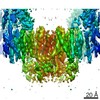 4515MC 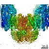 4432C 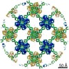 4439C 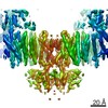 4441C 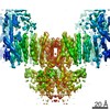 4513C 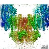 4514C 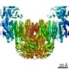 4516C  4517C 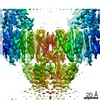 4518C 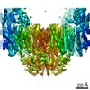 4519C 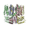 6i9dC 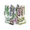 6iaxC 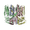 6qcyC 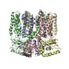 6qczC 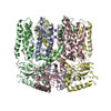 6qd1C 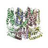 6qd2C 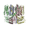 6qd3C 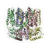 6qd4C C: 同じ文献を引用 ( M: このデータのモデリングに利用したマップデータ |
|---|---|
| 類似構造データ |


-リンク
 PDBj
PDBj


- 集合体
集合体
| 登録構造単位 | 
|
|---|---|
| 1 |
|
-要素
| #1: タンパク質 | 分子量: 37766.297 Da / 分子数: 4 / 由来タイプ: 組換発現 由来: (組換発現) Mesorhizobium loti MAFF303099 (根粒菌)発現宿主: #2: 化合物 |   分子量: 39.098 Da / 分子数: 2 / 由来タイプ: 合成 / 式: K 分子量: 39.098 Da / 分子数: 2 / 由来タイプ: 合成 / 式: K |
|---|
-実験情報
-実験
| 実験 | 手法: 電子顕微鏡法 |
|---|---|
| EM実験 | 試料の集合状態: 2D ARRAY / 3次元再構成法: 単粒子再構成法 |
- 試料調製
試料調製
| 構成要素 | 名称: MloK1 potassium channel in lipidic bilayer of 2D crystals タイプ: COMPLEX / Entity ID: #1 / 由来: RECOMBINANT |
|---|---|
| 分子量 | 値: 0.16 MDa / 実験値: YES |
| 由来(天然) | 生物種: Mesorhizobium loti MAFF303099 (根粒菌) |
| 由来(組換発現) | 生物種: |
| 緩衝液 | pH: 7.6 詳細: 20 mM KCl, 20 mM Tris-HCl pH 7.6, 1 mM BaCl2, 1 mM EDTA, 0.2 mM cAMP |
| 試料 | 包埋: NO / シャドウイング: NO / 染色: NO / 凍結: YES |
| 急速凍結 | 凍結剤: ETHANE |
- 電子顕微鏡撮影
| 実験機器 |  モデル: Titan Krios / 画像提供: FEI Company |
|---|---|
| 顕微鏡 | モデル: FEI TITAN KRIOS |
| 電子銃 | 電子線源:  FIELD EMISSION GUN / 加速電圧: 300 kV / 照射モード: OTHER FIELD EMISSION GUN / 加速電圧: 300 kV / 照射モード: OTHER |
| 電子レンズ | モード: BRIGHT FIELD / 倍率(公称値): 50000 X / 最大 デフォーカス(公称値): 4300 nm / 最小 デフォーカス(公称値): 750 nm / Cs: 2.7 mm |
| 試料ホルダ | 凍結剤: NITROGEN 試料ホルダーモデル: FEI TITAN KRIOS AUTOGRID HOLDER |
| 撮影 | 平均露光時間: 16 sec. / 電子線照射量: 45 e/Å2 / 検出モード: COUNTING フィルム・検出器のモデル: GATAN K2 SUMMIT (4k x 4k) 実像数: 270 詳細: The dataset of 346 movies recorded and processed for Kowal et al. (2018) 2 was employed. As reported, the data were collected on an FEI Titan Krios TEM equipped with a Gatan K2 DED. Total ...詳細: The dataset of 346 movies recorded and processed for Kowal et al. (2018) 2 was employed. As reported, the data were collected on an FEI Titan Krios TEM equipped with a Gatan K2 DED. Total dose: 40 e-/A^2 distributed over 40 movie frames. Pixel size: 1.3 A on the sample level (counting mode). Nominal tilt range: -55 to +55 degrees. |
- 解析
解析
| EMソフトウェア |
| ||||||||||||||||||||||||||||||||||||
|---|---|---|---|---|---|---|---|---|---|---|---|---|---|---|---|---|---|---|---|---|---|---|---|---|---|---|---|---|---|---|---|---|---|---|---|---|---|
| CTF補正 | 詳細: The defocus at the center of each particle box was estimated based on the tilt geometry. タイプ: PHASE FLIPPING AND AMPLITUDE CORRECTION | ||||||||||||||||||||||||||||||||||||
| 粒子像の選択 | 選択した粒子像数: 231688 詳細: The particle positions were given by the cross-correlation profile generated by the classical unbending algorithm. | ||||||||||||||||||||||||||||||||||||
| 対称性 | 点対称性: C4 (4回回転対称) | ||||||||||||||||||||||||||||||||||||
| 3次元再構成 | 解像度: 4.5 Å / 解像度の算出法: FSC 0.143 CUT-OFF / 粒子像の数: 31649 / アルゴリズム: FOURIER SPACE / クラス平均像の数: 1 / 対称性のタイプ: POINT | ||||||||||||||||||||||||||||||||||||
| 原子モデル構築 | プロトコル: FLEXIBLE FIT 詳細: The consensus model was flexibily fit in to the EM map of this class using Normal Mode Analysis with iMODFIT, and then real-space refined using PHENIX. | ||||||||||||||||||||||||||||||||||||
| 原子モデル構築 | PDB-ID: 6I9D Accession code: 6I9D / Source name: PDB / タイプ: experimental model |
