[English] 日本語
 Yorodumi
- PDB-6ne0: Structure of double-stranded target DNA engaged Csy complex from ... -
+ Open data
- Basic information
| Entry | Database: PDB / ID: 6ne0 | |||||||||
|---|---|---|---|---|---|---|---|---|---|---|
| Title | Structure of double-stranded target DNA engaged Csy complex from Pseudomonas aeruginosa (PA-14) | |||||||||
 Components Components |
| |||||||||
 Keywords Keywords | IMMUNE SYSTEM/RNA / type I-F CRISPR RNA-guided surveillance complex / viral protein mimic / Csy-complex / IMMUNE SYSTEM-RNA complex | |||||||||
| Function / homology |  Function and homology information Function and homology informationmaintenance of CRISPR repeat elements / endonuclease activity / defense response to virus / Hydrolases; Acting on ester bonds / RNA binding Similarity search - Function | |||||||||
| Biological species |  Pseudomonas aeruginosa UCBPP-PA14 (bacteria) Pseudomonas aeruginosa UCBPP-PA14 (bacteria) | |||||||||
| Method | ELECTRON MICROSCOPY / single particle reconstruction / cryo EM / Resolution: 3.4 Å | |||||||||
 Authors Authors | Chowdhury, S. / Rollins, M.F. / Carter, J. / Golden, S.M. / Miettinen, H.M. / Santiago-Frangos, A. / Faith, D. / Lawrence, M.C. / Wiedenheft, B. / Lander, G.C. | |||||||||
| Funding support |  United States, 2items United States, 2items
| |||||||||
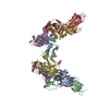
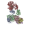
 Citation Citation |  Journal: Mol Cell / Year: 2019 Journal: Mol Cell / Year: 2019Title: Structure Reveals a Mechanism of CRISPR-RNA-Guided Nuclease Recruitment and Anti-CRISPR Viral Mimicry. Authors: MaryClare F Rollins / Saikat Chowdhury / Joshua Carter / Sarah M Golden / Heini M Miettinen / Andrew Santiago-Frangos / Dominick Faith / C Martin Lawrence / Gabriel C Lander / Blake Wiedenheft /  Abstract: Bacteria and archaea have evolved sophisticated adaptive immune systems that rely on CRISPR RNA (crRNA)-guided detection and nuclease-mediated elimination of invading nucleic acids. Here, we present ...Bacteria and archaea have evolved sophisticated adaptive immune systems that rely on CRISPR RNA (crRNA)-guided detection and nuclease-mediated elimination of invading nucleic acids. Here, we present the cryo-electron microscopy (cryo-EM) structure of the type I-F crRNA-guided surveillance complex (Csy complex) from Pseudomonas aeruginosa bound to a double-stranded DNA target. Comparison of this structure to previously determined structures of this complex reveals a ∼180-degree rotation of the C-terminal helical bundle on the "large" Cas8f subunit. We show that the double-stranded DNA (dsDNA)-induced conformational change in Cas8f exposes a Cas2/3 "nuclease recruitment helix" that is structurally homologous to a virally encoded anti-CRISPR protein (AcrIF3). Structural homology between Cas8f and AcrIF3 suggests that AcrIF3 is a mimic of the Cas8f nuclease recruitment helix. | |||||||||
| History |
|
- Structure visualization
| Movie |
 Movie viewer Movie viewer |
|---|---|
| Structure viewer | Molecule:  Molmil Molmil Jmol/JSmol Jmol/JSmol |
- Downloads & links
- Download
| PDBx/mmCIF format |  6ne0.cif.gz 6ne0.cif.gz | 543.8 KB | Display |  PDBx/mmCIF format PDBx/mmCIF format |
|---|---|---|---|---|
| PDB format |  pdb6ne0.ent.gz pdb6ne0.ent.gz | 434.4 KB | Display |  PDB format PDB format |
| PDBx/mmJSON format |  6ne0.json.gz 6ne0.json.gz | Tree view |  PDBx/mmJSON format PDBx/mmJSON format | |
| Others |  Other downloads Other downloads |
-Validation report
| Arichive directory |  https://data.pdbj.org/pub/pdb/validation_reports/ne/6ne0 https://data.pdbj.org/pub/pdb/validation_reports/ne/6ne0 ftp://data.pdbj.org/pub/pdb/validation_reports/ne/6ne0 ftp://data.pdbj.org/pub/pdb/validation_reports/ne/6ne0 | HTTPS FTP |
|---|
-Related structure data
| Related structure data |  9191MC M: map data used to model this data C: citing same article ( |
|---|---|
| Similar structure data |
- Links
Links
- Assembly
| Deposited unit | 
|
|---|---|
| 1 |
|
- Components
-CRISPR-associated protein ... , 3 types, 8 molecules ABCDEFGH
| #1: Protein | Mass: 49194.168 Da / Num. of mol.: 1 Source method: isolated from a genetically manipulated source Details: Gaps within the structure correspond to regions that could not be modeled due to missing map density. Source: (gene. exp.)  Pseudomonas aeruginosa UCBPP-PA14 (bacteria) Pseudomonas aeruginosa UCBPP-PA14 (bacteria)Strain: UCBPP-PA14 / Gene: csy1, PA14_33330 / Production host:  |
|---|---|
| #2: Protein | Mass: 36244.074 Da / Num. of mol.: 1 Source method: isolated from a genetically manipulated source Details: Gaps within the structure correspond to regions that could not be modeled due to missing map density. Source: (gene. exp.)  Pseudomonas aeruginosa UCBPP-PA14 (bacteria) Pseudomonas aeruginosa UCBPP-PA14 (bacteria)Strain: UCBPP-PA14 / Gene: csy2, PA14_33320 / Production host:  |
| #3: Protein | Mass: 37579.273 Da / Num. of mol.: 6 Source method: isolated from a genetically manipulated source Details: Gaps within the structure correspond to regions that could not be modeled due to missing map density. Source: (gene. exp.)  Pseudomonas aeruginosa UCBPP-PA14 (bacteria) Pseudomonas aeruginosa UCBPP-PA14 (bacteria)Strain: UCBPP-PA14 / Gene: csy3, csy1-3, PA14_33310 / Production host:  |
-DNA chain , 2 types, 2 molecules NO
| #6: DNA chain | Mass: 13584.703 Da / Num. of mol.: 1 / Source method: obtained synthetically Source: (synth.)  Pseudomonas aeruginosa UCBPP-PA14 (bacteria) Pseudomonas aeruginosa UCBPP-PA14 (bacteria) |
|---|---|
| #7: DNA chain | Mass: 10476.714 Da / Num. of mol.: 1 / Source method: obtained synthetically Details: Gaps within the structure correspond to regions that could not be modeled due to missing map density. Source: (synth.)  Pseudomonas aeruginosa UCBPP-PA14 (bacteria) Pseudomonas aeruginosa UCBPP-PA14 (bacteria) |
-Protein / RNA chain , 2 types, 2 molecules LM
| #4: Protein | Mass: 21675.781 Da / Num. of mol.: 1 Source method: isolated from a genetically manipulated source Source: (gene. exp.)  Pseudomonas aeruginosa UCBPP-PA14 (bacteria) Pseudomonas aeruginosa UCBPP-PA14 (bacteria)Strain: UCBPP-PA14 / Gene: cas6f, csy4, PA14_33300 / Production host:  References: UniProt: Q02MM2, Hydrolases; Acting on ester bonds |
|---|---|
| #5: RNA chain | Mass: 19265.404 Da / Num. of mol.: 1 / Source method: obtained synthetically Details: Gaps within the structure correspond to regions that could not be modeled due to missing map density. Source: (synth.)  Pseudomonas aeruginosa UCBPP-PA14 (bacteria) Pseudomonas aeruginosa UCBPP-PA14 (bacteria) |
-Experimental details
-Experiment
| Experiment | Method: ELECTRON MICROSCOPY |
|---|---|
| EM experiment | Aggregation state: PARTICLE / 3D reconstruction method: single particle reconstruction |
- Sample preparation
| Component | Name: DOUBLE-STRANDED TARGET DNA ENGAGED CSY COMPLEX FROM PSEUDOMONAS AERUGINOSA (PA-14) Type: COMPLEX / Entity ID: all / Source: RECOMBINANT | ||||||||||||||||||||||||||||||
|---|---|---|---|---|---|---|---|---|---|---|---|---|---|---|---|---|---|---|---|---|---|---|---|---|---|---|---|---|---|---|---|
| Molecular weight | Value: 0.36 MDa / Experimental value: NO | ||||||||||||||||||||||||||||||
| Source (natural) | Organism:  Pseudomonas aeruginosa UCBPP-PA14 (bacteria) Pseudomonas aeruginosa UCBPP-PA14 (bacteria) | ||||||||||||||||||||||||||||||
| Source (recombinant) | Organism:  | ||||||||||||||||||||||||||||||
| Buffer solution | pH: 7.5 | ||||||||||||||||||||||||||||||
| Buffer component |
| ||||||||||||||||||||||||||||||
| Specimen | Conc.: 2 mg/ml / Embedding applied: NO / Shadowing applied: NO / Staining applied: NO / Vitrification applied: YES | ||||||||||||||||||||||||||||||
| Specimen support | Grid material: GOLD / Grid mesh size: 300 divisions/in. / Grid type: UltrAuFoil | ||||||||||||||||||||||||||||||
| Vitrification | Instrument: HOMEMADE PLUNGER / Cryogen name: ETHANE Details: Freezing was carried out in a cold room at 4 degrees C and relative humidity of 98%. 5 ul sample was applied to plasma cleaned grid and manually blotted with Whatman 1 filter paper for 5-7 ...Details: Freezing was carried out in a cold room at 4 degrees C and relative humidity of 98%. 5 ul sample was applied to plasma cleaned grid and manually blotted with Whatman 1 filter paper for 5-7 sec before plunge freezing. |
- Electron microscopy imaging
| Experimental equipment |  Model: Talos Arctica / Image courtesy: FEI Company |
|---|---|
| Microscopy | Model: FEI TALOS ARCTICA Details: OBJECTIVE ASTIGMATISM WAS CORRECTED AT 36000X MAGNIFICATION USING THON RINGS VISUALIZED WITH A K2 CAMERA. |
| Electron gun | Electron source:  FIELD EMISSION GUN / Accelerating voltage: 200 kV / Illumination mode: FLOOD BEAM FIELD EMISSION GUN / Accelerating voltage: 200 kV / Illumination mode: FLOOD BEAM |
| Electron lens | Mode: BRIGHT FIELD / Nominal magnification: 36000 X / Nominal defocus max: 1500 nm / Nominal defocus min: 600 nm / Cs: 2.7 mm |
| Image recording | Electron dose: 0.58 e/Å2 / Film or detector model: GATAN K2 SUMMIT (4k x 4k) / Num. of real images: 3208 |
- Processing
| Software | Name: PHENIX / Version: dev_3304: / Classification: refinement | ||||||||||||||||||||
|---|---|---|---|---|---|---|---|---|---|---|---|---|---|---|---|---|---|---|---|---|---|
| EM software |
| ||||||||||||||||||||
| Image processing | Details: Super-resolution movie frames were Fourier-binned 2X2 times to a pixel size of 1.15 Angstrom/pixel prior to dose-weighted frame alignment using MotionCorr2. | ||||||||||||||||||||
| CTF correction | Type: PHASE FLIPPING AND AMPLITUDE CORRECTION | ||||||||||||||||||||
| Particle selection | Num. of particles selected: 1543677 Details: Particles were initially extracted binned by 2 with a box size of 144 pixels (2.3 Angstrom/pixel). The final processing was carried out with an unbinned particle stack with a box size of 288 ...Details: Particles were initially extracted binned by 2 with a box size of 144 pixels (2.3 Angstrom/pixel). The final processing was carried out with an unbinned particle stack with a box size of 288 pixels (1.15 Angstrom/pixel). | ||||||||||||||||||||
| Symmetry | Point symmetry: C1 (asymmetric) | ||||||||||||||||||||
| 3D reconstruction | Resolution: 3.4 Å / Resolution method: FSC 0.143 CUT-OFF / Num. of particles: 291227 / Algorithm: FOURIER SPACE Details: The final map was reconstructed using several focused maps from different sub-regions of the complex. These were initially aligned to each other and then stitched using the vop maximum ...Details: The final map was reconstructed using several focused maps from different sub-regions of the complex. These were initially aligned to each other and then stitched using the vop maximum function in UCSF chimera. The resolution of the final composite map is 3.2 angstrom. Symmetry type: POINT | ||||||||||||||||||||
| Atomic model building | Protocol: AB INITIO MODEL / Space: REAL Details: THE ATOMIC MODELS FOR CAS5F, CAS8F, CAS6F, AND CAS7F FROM THE CSY ACR COMPLEX (PDB ID 5UZ9) WERE USED AS INITIAL TEMPLATE MODELS FOR MODEL BUILDING. THESE WERE INDIVIDUALLY RIGID BODY-FITTED ...Details: THE ATOMIC MODELS FOR CAS5F, CAS8F, CAS6F, AND CAS7F FROM THE CSY ACR COMPLEX (PDB ID 5UZ9) WERE USED AS INITIAL TEMPLATE MODELS FOR MODEL BUILDING. THESE WERE INDIVIDUALLY RIGID BODY-FITTED INTO THE RECONSTRUCTED MAPS USING THE FIT MAP FUNCTION IN UCSF CHIMERA, AND RESIDUE REGISTERS AND BACKBONE GEOMETRIES WERE FIXED IN COOT. MODELS FOR THE CRRNA AND DNA STRANDS WERE ALSO MANUALLY BUILT INTO THE MAP USING COOT. | ||||||||||||||||||||
| Atomic model building | PDB-ID: 5UZ9 Accession code: 5UZ9 / Source name: PDB / Type: experimental model | ||||||||||||||||||||
| Refinement | Highest resolution: 3.4 Å |
 Movie
Movie Controller


 PDBj
PDBj


