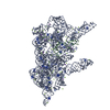
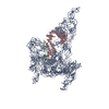
Movie
Movie Controller

[English] 日本語
 Yorodumi
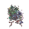
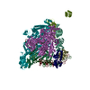
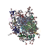
- PDB-6me0: Structure of a group II intron retroelement prior to DNA integration -
+ Open data

- Basic information
| Entry | Database: PDB / ID: 6me0 | |||||||||||||||
|---|---|---|---|---|---|---|---|---|---|---|---|---|---|---|---|---|
| Title | Structure of a group II intron retroelement prior to DNA integration | |||||||||||||||
 Components Components |
| |||||||||||||||
 Keywords Keywords | RNA/DNA/Nucleic Acid Binding protein / Group II intron / Retroelement / Retrotransposition / RNA-DNA-Nucleic Acid Binding protein complex | |||||||||||||||
| Function / homology |  Function and homology information Function and homology informationRNA-directed DNA polymerase activity / endonuclease activity / nucleic acid binding / zinc ion binding Similarity search - Function | |||||||||||||||
| Biological species |   Thermosynechococcus elongatus (bacteria) Thermosynechococcus elongatus (bacteria) | |||||||||||||||
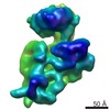
| Method | ELECTRON MICROSCOPY / single particle reconstruction / cryo EM / Resolution: 3.6 Å | |||||||||||||||
 Authors Authors | Haack, D. / Yan, X. / Zhang, C. / Hingey, J. / Lyumkis, D. / Baker, T.S. / Toor, N. | |||||||||||||||
| Funding support |  United States, 4items United States, 4items
| |||||||||||||||
 Citation Citation | Journal: Cell / Year: 2019 Title: Cryo-EM Structures of a Group II Intron Reverse Splicing into DNA. Authors: Daniel B Haack / Xiaodong Yan / Cheng Zhang / Jason Hingey / Dmitry Lyumkis / Timothy S Baker / Navtej Toor / Abstract: Group II introns are a class of retroelements that invade DNA through a copy-and-paste mechanism known as retrotransposition. Their coordinated activities occur within a complex that includes a ...Group II introns are a class of retroelements that invade DNA through a copy-and-paste mechanism known as retrotransposition. Their coordinated activities occur within a complex that includes a maturase protein, which promotes splicing through an unknown mechanism. The mechanism of splice site exchange within the RNA active site during catalysis also remains unclear. We determined two cryo-EM structures at 3.6-Å resolution of a group II intron reverse splicing into DNA. These structures reveal that the branch-site domain VI helix swings 90°, enabling substrate exchange during DNA integration. The maturase assists catalysis through a transient RNA-protein contact with domain VI that positions the branch-site adenosine for lariat formation during forward splicing. These findings provide the first direct evidence of the role the maturase plays during group II intron catalysis. The domain VI dynamics closely parallel spliceosomal branch-site helix movement and provide strong evidence for a retroelement origin of the spliceosome. | |||||||||||||||
| History |
|
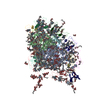
- Structure visualization
| Movie |
Movie viewer |
|---|---|
| Structure viewer | Molecule: MolmilJmol/JSmol |
- Downloads & links
-Download
| PDBx/mmCIF format | 6me0.cif.gz | 501 KB | Display | PDBx/mmCIF format |
|---|---|---|---|---|
| PDB format | pdb6me0.ent.gz | 373.8 KB | Display | PDB format |
| PDBx/mmJSON format | 6me0.json.gz | Tree view | PDBx/mmJSON format | |
| Others |  Other downloads Other downloads |
-Validation report
| Arichive directory | https://data.pdbj.org/pub/pdb/validation_reports/me/6me0ftp://data.pdbj.org/pub/pdb/validation_reports/me/6me0 | HTTPS FTP |
|---|
-Related structure data
| Related structure data | 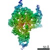 9105MC 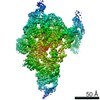 9106C 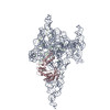 6mecC M: map data used to model this data C: citing same article ( |
|---|---|
| Similar structure data |

-Links
 PDBj
PDBj


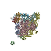
- Assembly
| Deposited unit | 
|
|---|---|
| 1 |
|
-Components
| #1: RNA chain | Mass: 280508.812 Da / Num. of mol.: 1 Source method: isolated from a genetically manipulated source Source: (gene. exp.) Thermosynechococcus elongatus (bacteria)Production host: | ||
|---|---|---|---|
| #2: DNA chain | Mass: 14032.991 Da / Num. of mol.: 1 / Source method: obtained synthetically / Source: (synth.) Thermosynechococcus elongatus (bacteria) | ||
| #3: Protein | Mass: 65065.121 Da / Num. of mol.: 1 Source method: isolated from a genetically manipulated source Source: (gene. exp.) Thermosynechococcus elongatus (strain BP-1) (bacteria)Strain: BP-1 / Gene: tll0114 / Plasmid: pET15b / Production host: | ||
| #4: Chemical | ChemComp-MG /   Mass: 24.305 Da / Num. of mol.: 32 / Source method: obtained synthetically / Formula: Mg Mass: 24.305 Da / Num. of mol.: 32 / Source method: obtained synthetically / Formula: Mg#5: Chemical | ChemComp-NA / |   Mass: 22.990 Da / Num. of mol.: 1 / Source method: obtained synthetically / Formula: Na Mass: 22.990 Da / Num. of mol.: 1 / Source method: obtained synthetically / Formula: Na |
-Experimental details
-Experiment
| Experiment | Method: ELECTRON MICROSCOPY |
|---|---|
| EM experiment | Aggregation state: PARTICLE / 3D reconstruction method: single particle reconstruction |
- Sample preparation
| Component | Name: T.el4h group II intron retroelement / Type: COMPLEX / Entity ID: #1-#3 / Source: RECOMBINANT |
|---|---|
| Molecular weight | Experimental value: NO |
| Source (natural) | Organism: Thermosynechococcus elongatus (bacteria) |
| Source (recombinant) | Organism: |
| Buffer solution | pH: 7.5 |
| Specimen | Conc.: 0.1 mg/ml / Embedding applied: NO / Shadowing applied: NO / Staining applied: NO / Vitrification applied: YES |
| Specimen support | Details: unspecified |
| Vitrification | Cryogen name: ETHANE |
- Electron microscopy imaging
| Experimental equipment |  Model: Titan Krios / Image courtesy: FEI Company |
|---|---|
| Microscopy | Model: FEI TITAN KRIOS |
| Electron gun | Electron source:  FIELD EMISSION GUN / Accelerating voltage: 300 kV / Illumination mode: FLOOD BEAM FIELD EMISSION GUN / Accelerating voltage: 300 kV / Illumination mode: FLOOD BEAM |
| Electron lens | Mode: BRIGHT FIELD |
| Image recording | Electron dose: 58 e/Å2 / Film or detector model: GATAN K2 SUMMIT (4k x 4k) |
- Processing
| Software |
| ||||||||||||||||||||||||
|---|---|---|---|---|---|---|---|---|---|---|---|---|---|---|---|---|---|---|---|---|---|---|---|---|---|
| EM software |
| ||||||||||||||||||||||||
| CTF correction | Type: PHASE FLIPPING AND AMPLITUDE CORRECTION | ||||||||||||||||||||||||
| Symmetry | Point symmetry: C1 (asymmetric) | ||||||||||||||||||||||||
| 3D reconstruction | Resolution: 3.6 Å / Resolution method: FSC 0.143 CUT-OFF / Num. of particles: 73144 / Num. of class averages: 2 / Symmetry type: POINT | ||||||||||||||||||||||||
| Atomic model building | PDB-ID: 4R0D Accession code: 4R0D / Source name: PDB / Type: experimental model | ||||||||||||||||||||||||
| Refinement | Stereochemistry target values: CDL v1.2 | ||||||||||||||||||||||||
| Refine LS restraints |
|