Movie
Movie Controller

[English] 日本語
 Yorodumi
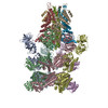
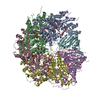
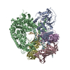
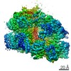
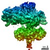
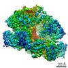
- PDB-6mdp: The D1 and D2 domain rings of NSF engaging the SNAP-25 N-terminus... -
+ Open data

- Basic information
| Entry | Database: PDB / ID: 6mdp | ||||||
|---|---|---|---|---|---|---|---|
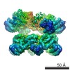
| Title | The D1 and D2 domain rings of NSF engaging the SNAP-25 N-terminus within the 20S supercomplex (focused refinement on D1/D2 rings, class 2) | ||||||
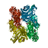
 Components Components |
| ||||||
 Keywords Keywords | HYDROLASE / SNARE / NSF / SNAP / ATPase / AAA / disassembly / synapse / membrane fusion / exocytosis | ||||||
| Function / homology |  Function and homology information Function and homology informationBLOC-1 complex / positive regulation of glutamate secretion, neurotransmission / synaptic vesicle fusion to presynaptic active zone membrane / Other interleukin signaling / synaptobrevin 2-SNAP-25-syntaxin-1a-complexin II complex / synaptobrevin 2-SNAP-25-syntaxin-1a complex / synaptobrevin 2-SNAP-25-syntaxin-1a-complexin I complex / presynaptic dense core vesicle exocytosis / extrinsic component of presynaptic membrane / calcium ion-regulated exocytosis of neurotransmitter ...BLOC-1 complex / positive regulation of glutamate secretion, neurotransmission / synaptic vesicle fusion to presynaptic active zone membrane / Other interleukin signaling / synaptobrevin 2-SNAP-25-syntaxin-1a-complexin II complex / synaptobrevin 2-SNAP-25-syntaxin-1a complex / synaptobrevin 2-SNAP-25-syntaxin-1a-complexin I complex / presynaptic dense core vesicle exocytosis / extrinsic component of presynaptic membrane / calcium ion-regulated exocytosis of neurotransmitter / Glutamate Neurotransmitter Release Cycle / Norepinephrine Neurotransmitter Release Cycle / Acetylcholine Neurotransmitter Release Cycle / Serotonin Neurotransmitter Release Cycle / GABA synthesis, release, reuptake and degradation / Dopamine Neurotransmitter Release Cycle / short-term synaptic potentiation / SNARE complex disassembly / regulation of establishment of protein localization / ribbon synapse / positive regulation of vesicle fusion / SNARE complex / SNAP receptor activity / ATP-dependent protein disaggregase activity / positive regulation of synaptic plasticity / intra-Golgi vesicle-mediated transport / positive regulation of hormone secretion / Golgi to plasma membrane protein transport / Golgi stack / neurotransmitter secretion / regulation of synaptic vesicle cycle / syntaxin-1 binding / Neutrophil degranulation / insulin secretion / vesicle-fusing ATPase / endosomal transport / myosin binding / regulation of synapse assembly / SNARE complex assembly / regulation of neuron projection development / synaptic vesicle priming / exocytosis / positive regulation of receptor recycling / associative learning / synaptic vesicle exocytosis / voltage-gated potassium channel activity / axonal growth cone / long-term memory / photoreceptor inner segment / somatodendritic compartment / voltage-gated potassium channel complex / axonogenesis / establishment of localization in cell / ionotropic glutamate receptor binding / SNARE binding / filopodium / PDZ domain binding / potassium ion transport / trans-Golgi network / locomotory behavior / intracellular protein transport / neuron differentiation / positive regulation of insulin secretion / calcium-dependent protein binding / terminal bouton / positive regulation of protein catabolic process / synaptic vesicle / long-term synaptic potentiation / lamellipodium / actin cytoskeleton / growth cone / presynaptic membrane / midbody / cell cortex / vesicle / cytoskeleton / transmembrane transporter binding / endosome / neuron projection / protein domain specific binding / axon / neuronal cell body / lipid binding / synapse / protein kinase binding / protein-containing complex binding / perinuclear region of cytoplasm / glutamatergic synapse / ATP hydrolysis activity / ATP binding / membrane / metal ion binding / identical protein binding / plasma membrane / cytoplasm / cytosol Similarity search - Function | ||||||
| Biological species |   Cricetulus griseus (Chinese hamster) Cricetulus griseus (Chinese hamster) | ||||||
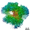
| Method | ELECTRON MICROSCOPY / single particle reconstruction / cryo EM / Resolution: 3.8 Å | ||||||
 Authors Authors | White, K.I. / Zhao, M. / Brunger, A.T. | ||||||
| Funding support |  United States, 1items United States, 1items
| ||||||
 Citation Citation | Journal: Elife / Year: 2018 Title: Structural principles of SNARE complex recognition by the AAA+ protein NSF. Authors: K Ian White / Minglei Zhao / Ucheor B Choi / Richard A Pfuetzner / Axel T Brunger / Abstract: The recycling of SNARE proteins following complex formation and membrane fusion is an essential process in eukaryotic trafficking. A highly conserved AAA+ protein, NSF (-ethylmaleimide sensitive ...The recycling of SNARE proteins following complex formation and membrane fusion is an essential process in eukaryotic trafficking. A highly conserved AAA+ protein, NSF (-ethylmaleimide sensitive factor) and an adaptor protein, SNAP (soluble NSF attachment protein), disassemble the SNARE complex. We report electron-cryomicroscopy structures of the complex of NSF, αSNAP, and the full-length soluble neuronal SNARE complex (composed of syntaxin-1A, synaptobrevin-2, SNAP-25A) in the presence of ATP under non-hydrolyzing conditions at ~3.9 Å resolution. These structures reveal electrostatic interactions by which two αSNAP molecules interface with a specific surface of the SNARE complex. This interaction positions the SNAREs such that the 15 N-terminal residues of SNAP-25A are loaded into the D1 ring pore of NSF via a spiral pattern of interactions between a conserved tyrosine NSF residue and SNAP-25A backbone atoms. This loading process likely precedes ATP hydrolysis. Subsequent ATP hydrolysis then drives complete disassembly. | ||||||
| History |
|
- Structure visualization
| Movie |
Movie viewer |
|---|---|
| Structure viewer | Molecule: MolmilJmol/JSmol |
- Downloads & links
-Download
| PDBx/mmCIF format | 6mdp.cif.gz | 959.1 KB | Display | PDBx/mmCIF format |
|---|---|---|---|---|
| PDB format | pdb6mdp.ent.gz | 788.2 KB | Display | PDB format |
| PDBx/mmJSON format | 6mdp.json.gz | Tree view | PDBx/mmJSON format | |
| Others |  Other downloads Other downloads |
-Validation report
| Arichive directory | https://data.pdbj.org/pub/pdb/validation_reports/md/6mdpftp://data.pdbj.org/pub/pdb/validation_reports/md/6mdp | HTTPS FTP |
|---|
-Related structure data
| Related structure data | 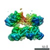 9103MC 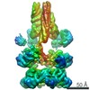 9100C 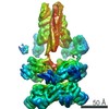 9101C 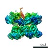 9102C 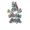 6mdmC 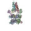 6mdnC 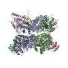 6mdoC M: map data used to model this data C: citing same article ( |
|---|---|
| Similar structure data |
-Links
 PDBj
PDBj


- Assembly
| Deposited unit | 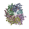
|
|---|---|
| 1 |
|
-Components
| #1: Protein | Mass: 85509.227 Da / Num. of mol.: 6 Source method: isolated from a genetically manipulated source Source: (gene. exp.) Cricetulus griseus (Chinese hamster) / Gene: NSF / Production host:  #2: Protein | | Mass: 23512.387 Da / Num. of mol.: 1 Source method: isolated from a genetically manipulated source Source: (gene. exp.) #3: Chemical |   Mass: 427.201 Da / Num. of mol.: 2 / Source method: obtained synthetically / Formula: C10H15N5O10P2 / Comment: ADP, energy-carrying molecule*YM Mass: 427.201 Da / Num. of mol.: 2 / Source method: obtained synthetically / Formula: C10H15N5O10P2 / Comment: ADP, energy-carrying molecule*YM#4: Chemical | ChemComp-ATP /   Mass: 507.181 Da / Num. of mol.: 9 / Source method: obtained synthetically / Formula: C10H16N5O13P3 / Comment: ATP, energy-carrying molecule*YM Mass: 507.181 Da / Num. of mol.: 9 / Source method: obtained synthetically / Formula: C10H16N5O13P3 / Comment: ATP, energy-carrying molecule*YM |
|---|
-Experimental details
-Experiment
| Experiment | Method: ELECTRON MICROSCOPY |
|---|---|
| EM experiment | Aggregation state: PARTICLE / 3D reconstruction method: single particle reconstruction |
- Sample preparation
| Component |
| |||||||||||||||||||||||||||||||||||
|---|---|---|---|---|---|---|---|---|---|---|---|---|---|---|---|---|---|---|---|---|---|---|---|---|---|---|---|---|---|---|---|---|---|---|---|---|
| Molecular weight |
| |||||||||||||||||||||||||||||||||||
| Source (natural) |
| |||||||||||||||||||||||||||||||||||
| Source (recombinant) |
| |||||||||||||||||||||||||||||||||||
| Buffer solution | pH: 8 | |||||||||||||||||||||||||||||||||||
| Buffer component |
| |||||||||||||||||||||||||||||||||||
| Specimen | Conc.: 15 mg/ml / Embedding applied: NO / Shadowing applied: NO / Staining applied: NO / Vitrification applied: YES | |||||||||||||||||||||||||||||||||||
| Specimen support | Grid material: COPPER / Grid mesh size: 400 divisions/in. / Grid type: Quantifoil R1.2/1.3 | |||||||||||||||||||||||||||||||||||
| Vitrification | Instrument: FEI VITROBOT MARK I / Cryogen name: ETHANE / Humidity: 100 % / Chamber temperature: 293 K / Details: Blot for 3.5 seconds before plunging. |
- Electron microscopy imaging
| Experimental equipment |  Model: Titan Krios / Image courtesy: FEI Company |
|---|---|
| Microscopy | Model: FEI TITAN KRIOS |
| Electron gun | Electron source:  FIELD EMISSION GUN / Accelerating voltage: 300 kV / Illumination mode: FLOOD BEAM FIELD EMISSION GUN / Accelerating voltage: 300 kV / Illumination mode: FLOOD BEAM |
| Electron lens | Mode: BRIGHT FIELD / Nominal defocus max: 3000 nm / Nominal defocus min: 1500 nm / Cs: 2.7 mm / C2 aperture diameter: 70 µm / Alignment procedure: COMA FREE |
| Specimen holder | Cryogen: NITROGEN / Specimen holder model: FEI TITAN KRIOS AUTOGRID HOLDER |
| Image recording | Average exposure time: 10 sec. / Electron dose: 58 e/Å2 / Detector mode: SUPER-RESOLUTION / Film or detector model: GATAN K2 SUMMIT (4k x 4k) / Num. of grids imaged: 2 / Num. of real images: 5418 |
| Image scans | Movie frames/image: 40 / Used frames/image: 2-40 |
- Processing
| EM software |
| ||||||||||||||||||||||||||||||||||||||||||||
|---|---|---|---|---|---|---|---|---|---|---|---|---|---|---|---|---|---|---|---|---|---|---|---|---|---|---|---|---|---|---|---|---|---|---|---|---|---|---|---|---|---|---|---|---|---|
| CTF correction | Details: CTF correction was carried out in Relion with reconstruction step. Type: PHASE FLIPPING AND AMPLITUDE CORRECTION | ||||||||||||||||||||||||||||||||||||||||||||
| Particle selection | Num. of particles selected: 475680 | ||||||||||||||||||||||||||||||||||||||||||||
| Symmetry | Point symmetry: C1 (asymmetric) | ||||||||||||||||||||||||||||||||||||||||||||
| 3D reconstruction | Resolution: 3.8 Å / Resolution method: FSC 0.143 CUT-OFF / Num. of particles: 184555 / Symmetry type: POINT | ||||||||||||||||||||||||||||||||||||||||||||
| Atomic model building | Protocol: RIGID BODY FIT / Space: REAL | ||||||||||||||||||||||||||||||||||||||||||||
| Atomic model building | PDB-ID: 3J96 Accession code: 3J96 / Source name: PDB / Type: experimental model |