 Movie
Movie Controller


+ Open data

- Basic information
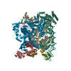
| Entry | Database: PDB / ID: 6jnx | ||||||
|---|---|---|---|---|---|---|---|
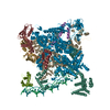
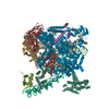
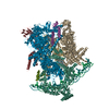
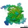
| Title | Cryo-EM structure of a Q-engaged arrested complex | ||||||
 Components Components |
| ||||||
 Keywords Keywords | TRANSCRIPTION / DNA / RNA / RNA polymerase / Antitermination | ||||||
| Function / homology |  Function and homology information Function and homology informationnegative regulation of termination of DNA-templated transcription / sigma factor antagonist complex / RNA polymerase complex / submerged biofilm formation / cellular response to cell envelope stress / bacterial-type RNA polymerase core enzyme binding / cytosolic DNA-directed RNA polymerase complex / regulation of DNA-templated transcription initiation / sigma factor activity / bacterial-type flagellum assembly ...negative regulation of termination of DNA-templated transcription / sigma factor antagonist complex / RNA polymerase complex / submerged biofilm formation / cellular response to cell envelope stress / bacterial-type RNA polymerase core enzyme binding / cytosolic DNA-directed RNA polymerase complex / regulation of DNA-templated transcription initiation / sigma factor activity / bacterial-type flagellum assembly / bacterial-type flagellum-dependent cell motility / nitrate assimilation / transcription elongation factor complex / regulation of DNA-templated transcription elongation / transcription antitermination / DNA-templated transcription initiation / cell motility / ribonucleoside binding / DNA-directed 5'-3' RNA polymerase activity / DNA-directed RNA polymerase / response to heat / protein-containing complex assembly / intracellular iron ion homeostasis / protein dimerization activity / response to antibiotic / negative regulation of DNA-templated transcription / magnesium ion binding / DNA binding / zinc ion binding / membrane / cytosol / cytoplasm Similarity search - Function | ||||||
| Biological species |   Enterobacteria phage SfI (virus)Enterobacteria phage P21 (virus) Enterobacteria phage SfI (virus)Enterobacteria phage P21 (virus) | ||||||
| Method | ELECTRON MICROSCOPY / single particle reconstruction / cryo EM / Resolution: 4.08 Å | ||||||
 Authors Authors | Feng, Y. / Shi, J. | ||||||
 Citation Citation | Journal: Nat Commun / Year: 2019 Title: Structural basis of Q-dependent transcription antitermination. Authors: Jing Shi / Xiang Gao / Tongguan Tian / Zhaoyang Yu / Bo Gao / Aijia Wen / Linlin You / Shenghai Chang / Xing Zhang / Yu Zhang / Yu Feng /  Abstract: Bacteriophage Q protein engages σ-dependent paused RNA polymerase (RNAP) by binding to a DNA site embedded in late gene promoter and renders RNAP resistant to termination signals. Here, we report a ...Bacteriophage Q protein engages σ-dependent paused RNA polymerase (RNAP) by binding to a DNA site embedded in late gene promoter and renders RNAP resistant to termination signals. Here, we report a single-particle cryo-electron microscopy (cryo-EM) structure of an intact Q-engaged arrested complex. The structure reveals key interactions responsible for σ-dependent pause, Q engagement, and Q-mediated transcription antitermination. The structure shows that two Q protomers (Q and Q) bind to a direct-repeat DNA site and contact distinct elements of the RNA exit channel. Notably, Q forms a narrow ring inside the RNA exit channel and renders RNAP resistant to termination signals by prohibiting RNA hairpin formation in the RNA exit channel. Because the RNA exit channel is conserved among all multisubunit RNAPs, it is likely to serve as an important contact site for regulators that modify the elongation properties of RNAP in other organisms, as well. | ||||||
| History |
|
- Structure visualization
| Movie |
Movie viewer |
|---|---|
| Structure viewer | Molecule: MolmilJmol/JSmol |
- Downloads & links
-Download
| PDBx/mmCIF format | 6jnx.cif.gz | 744.5 KB | Display | PDBx/mmCIF format |
|---|---|---|---|---|
| PDB format | pdb6jnx.ent.gz | 589.8 KB | Display | PDB format |
| PDBx/mmJSON format | 6jnx.json.gz | Tree view | PDBx/mmJSON format | |
| Others |  Other downloads Other downloads |
-Validation report
| Summary document | 6jnx_validation.pdf.gz | 1.1 MB | Display | wwPDB validaton report |
|---|---|---|---|---|
| Full document | 6jnx_full_validation.pdf.gz | 1.1 MB | Display | |
| Data in XML | 6jnx_validation.xml.gz | 103 KB | Display | |
| Data in CIF | 6jnx_validation.cif.gz | 160.8 KB | Display | |
| Arichive directory | https://data.pdbj.org/pub/pdb/validation_reports/jn/6jnxftp://data.pdbj.org/pub/pdb/validation_reports/jn/6jnx | HTTPS FTP |
-Related structure data
| Related structure data | 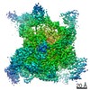 9852MC  6jnyC M: map data used to model this data C: citing same article ( |
|---|---|
| Similar structure data |


-Links
 PDBj
PDBj


- Assembly
| Deposited unit | 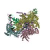
|
|---|---|
| 1 |
|
-Components
-DNA-directed RNA polymerase subunit ... , 4 types, 5 molecules ABCDE
| #1: Protein | Mass: 36558.680 Da / Num. of mol.: 2 Source method: isolated from a genetically manipulated source Source: (gene. exp.) #2: Protein | | Mass: 150820.875 Da / Num. of mol.: 1 Source method: isolated from a genetically manipulated source Source: (gene. exp.) #3: Protein | | Mass: 155366.781 Da / Num. of mol.: 1 Source method: isolated from a genetically manipulated source Source: (gene. exp.) #4: Protein | | Mass: 10249.547 Da / Num. of mol.: 1 Source method: isolated from a genetically manipulated source Source: (gene. exp.) |
|---|
-Protein , 2 types, 3 molecules FPQ
| #5: Protein | Mass: 70352.242 Da / Num. of mol.: 1 Source method: isolated from a genetically manipulated source Source: (gene. exp.) |
|---|---|
| #9: Protein | Mass: 18712.908 Da / Num. of mol.: 2 Source method: isolated from a genetically manipulated source Source: (gene. exp.) Enterobacteria phage SfI (virus) / Gene: Q / Production host: |
-DNA chain , 2 types, 2 molecules NT
| #6: DNA chain | Mass: 19558.576 Da / Num. of mol.: 1 / Source method: obtained synthetically / Source: (synth.) Enterobacteria phage P21 (virus) |
|---|---|
| #8: DNA chain | Mass: 19282.377 Da / Num. of mol.: 1 / Source method: obtained synthetically / Source: (synth.) Enterobacteria phage P21 (virus) |
-RNA chain , 1 types, 1 molecules R
| #7: RNA chain | Mass: 5893.548 Da / Num. of mol.: 1 / Source method: obtained synthetically / Source: (synth.) Enterobacteria phage P21 (virus) |
|---|
-Non-polymers , 2 types, 3 molecules 

| #10: Chemical | ChemComp-MG /  Mass: 24.305 Da / Num. of mol.: 1 / Source method: obtained synthetically / Formula: Mg Mass: 24.305 Da / Num. of mol.: 1 / Source method: obtained synthetically / Formula: Mg |
|---|---|
| #11: Chemical |  Mass: 65.409 Da / Num. of mol.: 2 / Source method: obtained synthetically / Formula: Zn Mass: 65.409 Da / Num. of mol.: 2 / Source method: obtained synthetically / Formula: Zn |
-Experimental details
-Experiment
| Experiment | Method: ELECTRON MICROSCOPY |
|---|---|
| EM experiment | Aggregation state: PARTICLE / 3D reconstruction method: single particle reconstruction |
- Sample preparation
| Component | Name: 21Q-engaged arrested complex / Type: COMPLEX / Entity ID: #1-#9 / Source: MULTIPLE SOURCES | ||||||||||||
|---|---|---|---|---|---|---|---|---|---|---|---|---|---|
| Molecular weight | Experimental value: NO | ||||||||||||
| Source (natural) |
| ||||||||||||
| Source (recombinant) | Organism: | ||||||||||||
| Buffer solution | pH: 8 | ||||||||||||
| Specimen | Conc.: 8 mg/ml / Embedding applied: NO / Shadowing applied: NO / Staining applied: NO / Vitrification applied: YES | ||||||||||||
| Vitrification | Cryogen name: ETHANE / Humidity: 95 % / Chamber temperature: 283 K |
- Electron microscopy imaging
| Experimental equipment |  Model: Titan Krios / Image courtesy: FEI Company |
|---|---|
| Microscopy | Model: FEI TITAN KRIOS |
| Electron gun | Electron source:  FIELD EMISSION GUN / Accelerating voltage: 300 kV / Illumination mode: FLOOD BEAM FIELD EMISSION GUN / Accelerating voltage: 300 kV / Illumination mode: FLOOD BEAM |
| Electron lens | Mode: BRIGHT FIELD |
| Image recording | Electron dose: 56 e/Å2 / Film or detector model: GATAN K2 SUMMIT (4k x 4k) |
- Processing
| CTF correction | Type: PHASE FLIPPING AND AMPLITUDE CORRECTION |
|---|---|
| Symmetry | Point symmetry: C1 (asymmetric) |
| 3D reconstruction | Resolution: 4.08 Å / Resolution method: FSC 0.143 CUT-OFF / Num. of particles: 64497 / Symmetry type: POINT |
