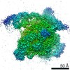
 Movie
Movie Controller

[English] 日本語
 Yorodumi
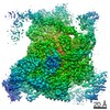
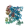
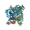
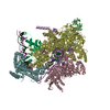
- PDB-7chw: Cryo-EM structure of an Escherichia coli RNAP-promoter open compl... -
+ Open data

- Basic information
| Entry | Database: PDB / ID: 7chw | ||||||
|---|---|---|---|---|---|---|---|
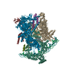
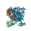
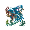
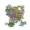
| Title | Cryo-EM structure of an Escherichia coli RNAP-promoter open complex (RPo) | ||||||
 Components Components |
| ||||||
 Keywords Keywords | TRANSCRIPTION/DNA / RNA polymerase / Transcription initiation complex / TRANSCRIPTION / TRANSCRIPTION-DNA complex | ||||||
| Function / homology |  Function and homology information Function and homology informationsigma factor antagonist complex / RNA polymerase complex / submerged biofilm formation / cellular response to cell envelope stress / regulation of DNA-templated transcription initiation / sigma factor activity / bacterial-type flagellum assembly / bacterial-type RNA polymerase core enzyme binding / cytosolic DNA-directed RNA polymerase complex / bacterial-type flagellum-dependent cell motility ...sigma factor antagonist complex / RNA polymerase complex / submerged biofilm formation / cellular response to cell envelope stress / regulation of DNA-templated transcription initiation / sigma factor activity / bacterial-type flagellum assembly / bacterial-type RNA polymerase core enzyme binding / cytosolic DNA-directed RNA polymerase complex / bacterial-type flagellum-dependent cell motility / nitrate assimilation / regulation of DNA-templated transcription elongation / transcription elongation factor complex / DNA-directed RNA polymerase complex / transcription antitermination / cell motility / DNA-templated transcription initiation / ribonucleoside binding / DNA-directed RNA polymerase / DNA-directed RNA polymerase activity / response to heat / protein-containing complex assembly / intracellular iron ion homeostasis / protein dimerization activity / transcription cis-regulatory region binding / response to antibiotic / negative regulation of DNA-templated transcription / regulation of DNA-templated transcription / DNA-templated transcription / magnesium ion binding / DNA binding / zinc ion binding / membrane / metal ion binding / cytoplasm / cytosol Similarity search - Function | ||||||
| Biological species |  | ||||||
| Method | ELECTRON MICROSCOPY / single particle reconstruction / Resolution: 3.58 Å | ||||||
 Authors Authors | Lin, W. / Feng, Y. | ||||||
 Citation Citation | Journal: Nucleic Acids Res / Year: 2020 Title: Structural basis for transcription inhibition by E. coli SspA. Authors: Fulin Wang / Jing Shi / Dingwei He / Bei Tong / Chao Zhang / Aijia Wen / Yu Zhang / Yu Feng / Wei Lin /  Abstract: Stringent starvation protein A (SspA) is an RNA polymerase (RNAP)-associated protein involved in nucleotide metabolism, acid tolerance and virulence of bacteria. Despite extensive biochemical and ...Stringent starvation protein A (SspA) is an RNA polymerase (RNAP)-associated protein involved in nucleotide metabolism, acid tolerance and virulence of bacteria. Despite extensive biochemical and genetic analyses, the precise regulatory role of SspA in transcription is still unknown, in part, because of a lack of structural information for bacterial RNAP in complex with SspA. Here, we report a 3.68 Å cryo-EM structure of an Escherichia coli RNAP-promoter open complex (RPo) with SspA. Unexpectedly, the structure reveals that SspA binds to the E. coli σ70-RNAP holoenzyme as a homodimer, interacting with σ70 region 4 and the zinc binding domain of EcoRNAP β' subunit simultaneously. Results from fluorescent polarization assays indicate the specific interactions between SspA and σ70 region 4 confer its σ selectivity, thereby avoiding its interactions with σs or other alternative σ factors. In addition, results from in vitro transcription assays verify that SspA inhibits transcription probably through suppressing promoter escape. Together, the results here provide a foundation for understanding the unique physiological function of SspA in transcription regulation in bacteria. | ||||||
| History |
|
- Structure visualization
| Movie |
Movie viewer |
|---|---|
| Structure viewer | Molecule: MolmilJmol/JSmol |
- Downloads & links
-Download
| PDBx/mmCIF format | 7chw.cif.gz | 725.9 KB | Display | PDBx/mmCIF format |
|---|---|---|---|---|
| PDB format | pdb7chw.ent.gz | 568.9 KB | Display | PDB format |
| PDBx/mmJSON format | 7chw.json.gz | Tree view | PDBx/mmJSON format | |
| Others |  Other downloads Other downloads |
-Validation report
| Arichive directory | https://data.pdbj.org/pub/pdb/validation_reports/ch/7chwftp://data.pdbj.org/pub/pdb/validation_reports/ch/7chw | HTTPS FTP |
|---|
-Related structure data
| Related structure data |  30376MC  7c97C M: map data used to model this data C: citing same article ( |
|---|---|
| Similar structure data |


-Links
 PDBj
PDBj


- Assembly
| Deposited unit | 
|
|---|---|
| 1 |
|
-Components
-DNA chain , 2 types, 2 molecules HG
| #1: DNA chain | Mass: 19671.666 Da / Num. of mol.: 1 / Source method: obtained synthetically / Source: (synth.) |
|---|---|
| #7: DNA chain | Mass: 19159.285 Da / Num. of mol.: 1 / Source method: obtained synthetically / Source: (synth.) |
-DNA-directed RNA polymerase subunit ... , 4 types, 6 molecules KABCDE
| #2: Protein | Mass: 36558.680 Da / Num. of mol.: 3 Source method: isolated from a genetically manipulated source Source: (gene. exp.) References: UniProt: U9ZUN7, UniProt: P0A7Z4*PLUS, DNA-directed RNA polymerase #3: Protein | | Mass: 150804.922 Da / Num. of mol.: 1 Source method: isolated from a genetically manipulated source Source: (gene. exp.) Strain: K12 Gene: rpoB, groN, nitB, rif, ron, stl, stv, tabD, b3987, JW3950 Production host: #4: Protein | | Mass: 155366.781 Da / Num. of mol.: 1 Source method: isolated from a genetically manipulated source Source: (gene. exp.) References: UniProt: D7Y6A2, UniProt: P0A8T7*PLUS, DNA-directed RNA polymerase #5: Protein | | Mass: 10249.547 Da / Num. of mol.: 1 Source method: isolated from a genetically manipulated source Source: (gene. exp.) References: UniProt: A0A070UPX4, UniProt: P0A800*PLUS, DNA-directed RNA polymerase |
|---|
-Protein , 1 types, 1 molecules F
| #6: Protein | Mass: 70352.242 Da / Num. of mol.: 1 Source method: isolated from a genetically manipulated source Source: (gene. exp.) |
|---|
-Non-polymers , 2 types, 3 molecules 

| #8: Chemical | ChemComp-MG /  Mass: 24.305 Da / Num. of mol.: 1 / Source method: obtained synthetically / Formula: Mg / Feature type: SUBJECT OF INVESTIGATION Mass: 24.305 Da / Num. of mol.: 1 / Source method: obtained synthetically / Formula: Mg / Feature type: SUBJECT OF INVESTIGATION |
|---|---|
| #9: Chemical |  Mass: 65.409 Da / Num. of mol.: 2 / Source method: obtained synthetically / Formula: Zn / Feature type: SUBJECT OF INVESTIGATION Mass: 65.409 Da / Num. of mol.: 2 / Source method: obtained synthetically / Formula: Zn / Feature type: SUBJECT OF INVESTIGATION |
-Details
| Has ligand of interest | Y |
|---|
-Experimental details
-Experiment
| Experiment | Method: ELECTRON MICROSCOPY |
|---|---|
| EM experiment | Aggregation state: PARTICLE / 3D reconstruction method: single particle reconstruction |
- Sample preparation
| Component | Name: Escherichia coli RNAP-promoter open complex (RPo) / Type: COMPLEX / Entity ID: #1-#7 / Source: RECOMBINANT |
|---|---|
| Molecular weight | Value: 0.45 kDa/nm / Experimental value: YES |
| Source (natural) | Organism: |
| Source (recombinant) | Organism: |
| Buffer solution | pH: 7.9 |
| Specimen | Embedding applied: NO / Shadowing applied: NO / Staining applied: NO / Vitrification applied: NO |
- Electron microscopy imaging
| Experimental equipment |  Model: Titan Krios / Image courtesy: FEI Company |
|---|---|
| Microscopy | Model: FEI TITAN KRIOS |
| Electron gun | Electron source:  FIELD EMISSION GUN / Accelerating voltage: 300 kV / Illumination mode: FLOOD BEAM FIELD EMISSION GUN / Accelerating voltage: 300 kV / Illumination mode: FLOOD BEAM |
| Electron lens | Mode: BRIGHT FIELD |
| Image recording | Electron dose: 59 e/Å2 / Detector mode: COUNTING / Film or detector model: GATAN K2 SUMMIT (4k x 4k) |
- Processing
| EM software |
| ||||||||||||
|---|---|---|---|---|---|---|---|---|---|---|---|---|---|
| CTF correction | Type: PHASE FLIPPING AND AMPLITUDE CORRECTION | ||||||||||||
| 3D reconstruction | Resolution: 3.58 Å / Resolution method: FSC 0.143 CUT-OFF / Num. of particles: 199208 / Symmetry type: POINT |
