 Movie
Movie Controller

[English] 日本語
 Yorodumi
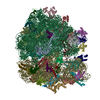
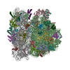
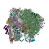
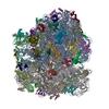
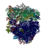
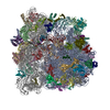
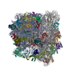
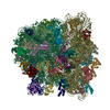
- PDB-6h4n: Structure of a hibernating 100S ribosome reveals an inactive conf... -
+ Open data

- Basic information
| Entry | Database: PDB / ID: 6h4n | |||||||||
|---|---|---|---|---|---|---|---|---|---|---|
| Title | Structure of a hibernating 100S ribosome reveals an inactive conformation of the ribosomal protein S1 - 70S Hibernating E. coli Ribosome | |||||||||
 Components Components |
| |||||||||
 Keywords Keywords | RIBOSOME / 100S / cryo-EM / E-site tRNA / hibernation / HPF / RMF / S1 | |||||||||
| Function / homology |  Function and homology information Function and homology informationdormancy process / negative regulation of translation in response to stress / negative regulation of translational elongation / positive regulation of cytoplasmic translation / negative regulation of cytoplasmic translational initiation / cellular response to stress / ornithine decarboxylase inhibitor activity / transcription antitermination factor activity, RNA binding / misfolded RNA binding / Group I intron splicing ...dormancy process / negative regulation of translation in response to stress / negative regulation of translational elongation / positive regulation of cytoplasmic translation / negative regulation of cytoplasmic translational initiation / cellular response to stress / ornithine decarboxylase inhibitor activity / transcription antitermination factor activity, RNA binding / misfolded RNA binding / Group I intron splicing / ribosomal small subunit binding / RNA folding / transcriptional attenuation / positive regulation of ribosome biogenesis / endoribonuclease inhibitor activity / RNA-binding transcription regulator activity / negative regulation of cytoplasmic translation / four-way junction DNA binding / DnaA-L2 complex / translation repressor activity / negative regulation of translational initiation / regulation of mRNA stability / negative regulation of DNA-templated DNA replication initiation / mRNA regulatory element binding translation repressor activity / assembly of large subunit precursor of preribosome / positive regulation of RNA splicing / ribosome assembly / transcription elongation factor complex / regulation of DNA-templated transcription elongation / cytosolic ribosome assembly / response to reactive oxygen species / DNA endonuclease activity / transcription antitermination / translational initiation / regulation of cell growth / DNA-templated transcription termination / response to radiation / maintenance of translational fidelity / mRNA 5'-UTR binding / ribosome biogenesis / large ribosomal subunit / regulation of translation / transferase activity / ribosome binding / ribosomal small subunit biogenesis / ribosomal small subunit assembly / small ribosomal subunit / 5S rRNA binding / ribosomal large subunit assembly / cytosolic small ribosomal subunit / large ribosomal subunit rRNA binding / small ribosomal subunit rRNA binding / cytosolic large ribosomal subunit / cytoplasmic translation / tRNA binding / single-stranded RNA binding / negative regulation of translation / rRNA binding / structural constituent of ribosome / ribosome / translation / response to antibiotic / negative regulation of DNA-templated transcription / mRNA binding / DNA binding / RNA binding / zinc ion binding / membrane / cytosol / cytoplasm Similarity search - Function | |||||||||
| Biological species |  | |||||||||
| Method | ELECTRON MICROSCOPY / single particle reconstruction / cryo EM / Resolution: 3 Å | |||||||||
 Authors Authors | Beckert, B. / Turk, M. / Czech, A. / Berninghausen, O. / Beckmann, R. / Ignatova, Z. / Plitzko, J. / Wilson, N.D. | |||||||||
| Funding support |  Germany, 2items Germany, 2items
| |||||||||
 Citation Citation | Journal: Nat Microbiol / Year: 2018 Title: Structure of a hibernating 100S ribosome reveals an inactive conformation of the ribosomal protein S1. Authors: Bertrand Beckert / Martin Turk / Andreas Czech / Otto Berninghausen / Roland Beckmann / Zoya Ignatova / Jürgen M Plitzko / Daniel N Wilson / Abstract: To survive under conditions of stress, such as nutrient deprivation, bacterial 70S ribosomes dimerize to form hibernating 100S particles. In γ-proteobacteria, such as Escherichia coli, 100S ...To survive under conditions of stress, such as nutrient deprivation, bacterial 70S ribosomes dimerize to form hibernating 100S particles. In γ-proteobacteria, such as Escherichia coli, 100S formation requires the ribosome modulation factor (RMF) and the hibernation promoting factor (HPF). Here we present single-particle cryo-electron microscopy structures of hibernating 70S and 100S particles isolated from stationary-phase E. coli cells at 3.0 Å and 7.9 Å resolution, respectively. The structures reveal the binding sites for HPF and RMF as well as the unexpected presence of deacylated E-site transfer RNA and ribosomal protein bS1. HPF interacts with the anticodon-stem-loop of the E-tRNA and occludes the binding site for the messenger RNA as well as A- and P-site tRNAs. RMF facilitates stabilization of a compact conformation of bS1, which together sequester the anti-Shine-Dalgarno sequence of the 16S ribosomal RNA (rRNA), thereby inhibiting translation initiation. At the dimerization interface, the C-terminus of uS2 probes the mRNA entrance channel of the symmetry-related particle, thus suggesting that dimerization inactivates ribosomes by blocking the binding of mRNA within the channel. The back-to-back E. coli 100S arrangement is distinct from 100S particles observed previously in Gram-positive bacteria, and reveals a unique role for bS1 in translation regulation. | |||||||||
| History |
|
- Structure visualization
| Movie |
Movie viewer |
|---|---|
| Structure viewer | Molecule: MolmilJmol/JSmol |
- Downloads & links
-Download
| PDBx/mmCIF format | 6h4n.cif.gz | 3.2 MB | Display | PDBx/mmCIF format |
|---|---|---|---|---|
| PDB format | pdb6h4n.ent.gz | Display | PDB format | |
| PDBx/mmJSON format | 6h4n.json.gz | Tree view | PDBx/mmJSON format | |
| Others |  Other downloads Other downloads |
-Validation report
| Summary document | 6h4n_validation.pdf.gz | 1.3 MB | Display | wwPDB validaton report |
|---|---|---|---|---|
| Full document | 6h4n_full_validation.pdf.gz | 1.4 MB | Display | |
| Data in XML | 6h4n_validation.xml.gz | 213.3 KB | Display | |
| Data in CIF | 6h4n_validation.cif.gz | 376.8 KB | Display | |
| Arichive directory | https://data.pdbj.org/pub/pdb/validation_reports/h4/6h4nftp://data.pdbj.org/pub/pdb/validation_reports/h4/6h4n | HTTPS FTP |
-Related structure data
| Related structure data | 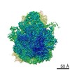 0137MC 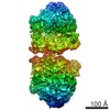 0139C  6h58C M: map data used to model this data C: citing same article ( |
|---|---|
| Similar structure data |
-Links
 PDBj
PDBj


- Assembly
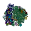
| Deposited unit | 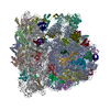
|
|---|---|
| 1 |
|
-Components
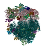
-RNA chain , 5 types, 5 molecules aABwI
| #1: RNA chain | Mass: 497197.531 Da / Num. of mol.: 1 / Source method: isolated from a natural source / Source: (natural) |
|---|---|
| #22: RNA chain | Mass: 941305.250 Da / Num. of mol.: 1 / Source method: isolated from a natural source / Source: (natural) |
| #23: RNA chain | Mass: 38813.133 Da / Num. of mol.: 1 / Source method: isolated from a natural source / Source: (natural) |
| #53: RNA chain | Mass: 24771.770 Da / Num. of mol.: 1 / Source method: isolated from a natural source / Source: (natural) |
| #57: RNA chain | Mass: 1482.920 Da / Num. of mol.: 1 / Source method: isolated from a natural source / Source: (natural) |
+30S ribosomal protein ... , 21 types, 21 molecules bcdefghijklmnopqrstuy
+50S ribosomal protein ... , 29 types, 29 molecules CDEFGHJKLMNOPQRSTUVWXYZ012346
-Protein , 2 types, 2 molecules vx
| #54: Protein | Mass: 6520.523 Da / Num. of mol.: 1 / Source method: isolated from a natural source / Source: (natural) |
|---|---|
| #56: Protein | Mass: 10768.230 Da / Num. of mol.: 1 / Source method: isolated from a natural source / Source: (natural) |
-Details
| Has protein modification | Y |
|---|
-Experimental details
-Experiment
| Experiment | Method: ELECTRON MICROSCOPY |
|---|---|
| EM experiment | Aggregation state: PARTICLE / 3D reconstruction method: single particle reconstruction |
- Sample preparation
| Component | Name: Structure of a hibernating 100S ribosome reveals an inactive conformation of the ribosomal protein S1 Type: RIBOSOME Details: Here we present single particle cryo-electron microscopy structures of hibernating 70S and 100S particles isolated from stationary phase E. coli cells at 3.0 and 7.9 resolution, respectively. ...Details: Here we present single particle cryo-electron microscopy structures of hibernating 70S and 100S particles isolated from stationary phase E. coli cells at 3.0 and 7.9 resolution, respectively. The structures reveal the binding sites for HPF and RMF as well as the unexpected presence of deacylated E-site tRNA and ribosomal protein bS1. HPF interacts with the anticodon-stem-loop of the E-tRNA and occludes the binding site for the mRNA as well as A- and P-site tRNAs. RMF facilitates stabilization of a compact conformation of bS1, which together sequester the anti-Shine-Dalgarno sequence of the 16S ribosomal RNA (rRNA), thereby inhibiting translation initiation. At the dimerization interface, the C-terminus of uS2 probes the mRNA entrance channel of the symmetry related particle, thus suggesting that dimerization inactivates ribosomes by blocking the binding of mRNA within the channel. Entity ID: all / Source: NATURAL |
|---|---|
| Molecular weight | Units: MEGADALTONS / Experimental value: NO |
| Source (natural) | Organism: |
| Buffer solution | pH: 7.5 Details: B100S buffer (25 mM Hepes pH 7.5, 100 mM KOAc, 15 mM Mg(OAc)2, 1 mM dithiothreitol) |
| Specimen | Embedding applied: NO / Shadowing applied: NO / Staining applied: NO / Vitrification applied: YES |
| Specimen support | Grid material: GOLD / Grid type: Quantifoil, UltrAuFoil, R1.2/1.3 |
| Vitrification | Instrument: FEI VITROBOT MARK III / Cryogen name: ETHANE-PROPANE |
- Electron microscopy imaging
| Experimental equipment |  Model: Titan Krios / Image courtesy: FEI Company |
|---|---|
| Microscopy | Model: FEI TITAN KRIOS |
| Electron gun | Electron source:  FIELD EMISSION GUN / Accelerating voltage: 300 kV / Illumination mode: SPOT SCAN FIELD EMISSION GUN / Accelerating voltage: 300 kV / Illumination mode: SPOT SCAN |
| Electron lens | Mode: BRIGHT FIELD |
| Image recording | Electron dose: 2.5 e/Å2 / Film or detector model: FEI FALCON II (4k x 4k) |
- Processing
| EM software |
| ||||||||||||||||||||||||
|---|---|---|---|---|---|---|---|---|---|---|---|---|---|---|---|---|---|---|---|---|---|---|---|---|---|
| CTF correction | Type: PHASE FLIPPING AND AMPLITUDE CORRECTION | ||||||||||||||||||||||||
| Symmetry | Point symmetry: C1 (asymmetric) | ||||||||||||||||||||||||
| 3D reconstruction | Resolution: 3 Å / Resolution method: FSC 0.143 CUT-OFF / Num. of particles: 188304 / Symmetry type: POINT | ||||||||||||||||||||||||
| Atomic model building | Protocol: RIGID BODY FIT |
