 Movie
Movie Controller

[English] 日本語
 Yorodumi
- PDB-6gyb: Cryo-EM structure of the bacteria-killing type IV secretion syste... -
+ Open data

- Basic information
| Entry | Database: PDB / ID: 6gyb | ||||||||||||
|---|---|---|---|---|---|---|---|---|---|---|---|---|---|
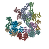
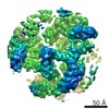
| Title | Cryo-EM structure of the bacteria-killing type IV secretion system core complex from Xanthomonas citri | ||||||||||||
 Components Components |
| ||||||||||||
 Keywords Keywords | MEMBRANE PROTEIN / core complex / bacterial killing / protein transport / bacterial Type IV Secretion System | ||||||||||||
| Function / homology |  Function and homology information Function and homology information | ||||||||||||
| Biological species |  Xanthomonas axonopodis pv. citri (bacteria) Xanthomonas axonopodis pv. citri (bacteria) | ||||||||||||
| Method | ELECTRON MICROSCOPY / single particle reconstruction / cryo EM / Resolution: 3.28 Å | ||||||||||||
 Authors Authors | Sgro, G.G. / Costa, T.R.D. / Farah, C.S. / Waksman, G. | ||||||||||||
| Funding support |  United Kingdom, United Kingdom,  Brazil, 3items Brazil, 3items
| ||||||||||||
 Citation Citation | Journal: Nat Microbiol / Year: 2018 Title: Cryo-EM structure of the bacteria-killing type IV secretion system core complex from Xanthomonas citri. Authors: Germán G Sgro / Tiago R D Costa / William Cenens / Diorge P Souza / Alexandre Cassago / Luciana Coutinho de Oliveira / Roberto K Salinas / Rodrigo V Portugal / Chuck S Farah / Gabriel Waksman /  Abstract: Type IV secretion (T4S) systems form the most common and versatile class of secretion systems in bacteria, capable of injecting both proteins and DNAs into host cells. T4S systems are typically ...Type IV secretion (T4S) systems form the most common and versatile class of secretion systems in bacteria, capable of injecting both proteins and DNAs into host cells. T4S systems are typically composed of 12 components that form 2 major assemblies: the inner membrane complex embedded in the inner membrane and the core complex embedded in both the inner and outer membranes. Here we present the 3.3 Å-resolution cryo-electron microscopy model of the T4S system core complex from Xanthomonas citri, a phytopathogen that utilizes this system to kill bacterial competitors. An extensive mutational investigation was performed to probe the vast network of protein-protein interactions in this 1.13-MDa assembly. This structure expands our knowledge of the molecular details of T4S system organization, assembly and evolution. | ||||||||||||
| History |
|
- Structure visualization
| Movie |
Movie viewer |
|---|---|
| Structure viewer | Molecule: MolmilJmol/JSmol |
- Downloads & links
-Download
| PDBx/mmCIF format | 6gyb.cif.gz | 1.3 MB | Display | PDBx/mmCIF format |
|---|---|---|---|---|
| PDB format | pdb6gyb.ent.gz | 1.1 MB | Display | PDB format |
| PDBx/mmJSON format | 6gyb.json.gz | Tree view | PDBx/mmJSON format | |
| Others |  Other downloads Other downloads |
-Validation report
| Arichive directory | https://data.pdbj.org/pub/pdb/validation_reports/gy/6gybftp://data.pdbj.org/pub/pdb/validation_reports/gy/6gyb | HTTPS FTP |
|---|
-Related structure data
| Related structure data | 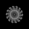 0089MC M: map data used to model this data C: citing same article ( |
|---|---|
| Similar structure data | |
| Other databases |
|


-Links
 PDBj
PDBj- Assembly
| Deposited unit | 
|
|---|---|
| 1 |
|
-Components
| #1: Protein | Mass: 14762.795 Da / Num. of mol.: 14 Source method: isolated from a genetically manipulated source Source: (gene. exp.) Xanthomonas axonopodis pv. citri (strain 306) (bacteria)Gene: XAC2622 / Production host: #2: Protein | Mass: 29359.385 Da / Num. of mol.: 14 Source method: isolated from a genetically manipulated source Source: (gene. exp.) Xanthomonas axonopodis pv. citri (strain 306) (bacteria)Strain: 306 / Gene: virB9, XAC2620 / Production host: #3: Protein | Mass: 43392.469 Da / Num. of mol.: 14 Source method: isolated from a genetically manipulated source Source: (gene. exp.) Xanthomonas axonopodis pv. citri (strain 306) (bacteria)Strain: 306 / Gene: virB10, XAC2619 / Production host: Has protein modification | Y | |
|---|
-Experimental details
-Experiment
| Experiment | Method: ELECTRON MICROSCOPY |
|---|---|
| EM experiment | Aggregation state: PARTICLE / 3D reconstruction method: single particle reconstruction |
- Sample preparation
| Component | Name: Core complex of a bacterial killing type IV secretion system from Xanthomonas Type: COMPLEX Details: Fourteen copies of each of the following three subunits: VirB7, VirB9 and VirB10 Entity ID: all / Source: RECOMBINANT | ||||||||||||||||||||
|---|---|---|---|---|---|---|---|---|---|---|---|---|---|---|---|---|---|---|---|---|---|
| Molecular weight | Experimental value: NO | ||||||||||||||||||||
| Source (natural) | Organism: Xanthomonas axonopodis pv. citri str. 306 (bacteria) | ||||||||||||||||||||
| Source (recombinant) | Organism: | ||||||||||||||||||||
| Buffer solution | pH: 8 | ||||||||||||||||||||
| Buffer component |
| ||||||||||||||||||||
| Specimen | Conc.: 0.3 mg/ml / Embedding applied: NO / Shadowing applied: NO / Staining applied: NO / Vitrification applied: YES | ||||||||||||||||||||
| Vitrification | Instrument: FEI VITROBOT MARK IV / Cryogen name: ETHANE / Humidity: 100 % / Chamber temperature: 295.2 K Details: Blot for 4.5 seconds after 30 seconds of incubation. |
- Electron microscopy imaging
| Experimental equipment |  Model: Titan Krios / Image courtesy: FEI Company |
|---|---|
| Microscopy | Model: FEI TITAN KRIOS |
| Electron gun | Electron source:  FIELD EMISSION GUN / Accelerating voltage: 300 kV / Illumination mode: FLOOD BEAM FIELD EMISSION GUN / Accelerating voltage: 300 kV / Illumination mode: FLOOD BEAM |
| Electron lens | Mode: BRIGHT FIELD |
| Image recording | Average exposure time: 12 sec. / Electron dose: 60 e/Å2 / Detector mode: SUPER-RESOLUTION / Film or detector model: GATAN K2 QUANTUM (4k x 4k) / Num. of grids imaged: 1 / Num. of real images: 1469 |
| Image scans | Movie frames/image: 40 |
- Processing
| Software | Name: PHENIX / Version: 1.12_2829: / Classification: refinement | ||||||||||||||||||||||||||||||||||||||||||||
|---|---|---|---|---|---|---|---|---|---|---|---|---|---|---|---|---|---|---|---|---|---|---|---|---|---|---|---|---|---|---|---|---|---|---|---|---|---|---|---|---|---|---|---|---|---|
| EM software |
| ||||||||||||||||||||||||||||||||||||||||||||
| CTF correction | Type: PHASE FLIPPING AND AMPLITUDE CORRECTION | ||||||||||||||||||||||||||||||||||||||||||||
| Particle selection | Num. of particles selected: 185079 | ||||||||||||||||||||||||||||||||||||||||||||
| Symmetry | Point symmetry: C14 (14 fold cyclic) | ||||||||||||||||||||||||||||||||||||||||||||
| 3D reconstruction | Resolution: 3.28 Å / Resolution method: FSC 0.143 CUT-OFF / Num. of particles: 142306 / Algorithm: BACK PROJECTION / Num. of class averages: 1 / Symmetry type: POINT | ||||||||||||||||||||||||||||||||||||||||||||
| Atomic model building | B value: 138 / Space: REAL / Target criteria: Cross-correlation coefficient Details: The electron density was clearly interpretable, which allowed us to build a de novo structural model. This process began by fitting the crystallographic model of the X. citri VirB7 C- ...Details: The electron density was clearly interpretable, which allowed us to build a de novo structural model. This process began by fitting the crystallographic model of the X. citri VirB7 C-terminal N0 domain (PDB:3OV5) and the NMR model of the X. citri VirB9CTD-VirB7NTD complex (PDB:2N01) in order to identify the map with the correct handedness. Models were positioned using Fit in map tool in Chimera, and saved relative to the map. Using these as starting points, we were able to manually build the rest of the model for VirB7 and VirB9CTD, and the de novo models for VirB10CTD, VirB10NTD_150-161 and VirB9NTD using Coot. In this manner, we obtained a combined model for a single VirB7-VirB9-VirB10 heterotrimer unit, which was submitted to iterative rounds of real space refinement and building using PHENIX and Coot software, respectively. Thirteen more copies of the refined heterotrimer were then fit into the density map using Chimera and new rounds of real space refinement (now using NCS for the 42 chains contained in the structure) and building using PHENIX and Coot, respectively, were executed until we obtained good parameters for Ramachandran plot and MolProbity. Chimera and PyMol were used for map and model visualization and figure production. | ||||||||||||||||||||||||||||||||||||||||||||
| Refine LS restraints |
|
