+ データを開く
- 基本情報
基本情報
| 登録情報 | データベース: PDB / ID: 6gv4 | |||||||||||||||
|---|---|---|---|---|---|---|---|---|---|---|---|---|---|---|---|---|
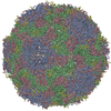
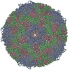
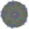
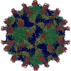
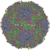
| タイトル | High-resolution Cryo-EM of Fab-labeled human parechovirus 3 | |||||||||||||||
 要素 要素 |
| |||||||||||||||
 キーワード キーワード | VIRUS / human parechovirus / antibody / RNA | |||||||||||||||
| 機能・相同性 |  機能・相同性情報 機能・相同性情報T=pseudo3 icosahedral viral capsid / host cell cytoplasmic vesicle membrane / cytoplasmic vesicle membrane / channel activity / monoatomic ion transmembrane transport / RNA helicase activity / symbiont entry into host cell / viral RNA genome replication / cysteine-type endopeptidase activity / RNA-dependent RNA polymerase activity ...T=pseudo3 icosahedral viral capsid / host cell cytoplasmic vesicle membrane / cytoplasmic vesicle membrane / channel activity / monoatomic ion transmembrane transport / RNA helicase activity / symbiont entry into host cell / viral RNA genome replication / cysteine-type endopeptidase activity / RNA-dependent RNA polymerase activity / DNA-templated transcription / virion attachment to host cell / structural molecule activity / proteolysis / RNA binding / ATP binding 類似検索 - 分子機能 | |||||||||||||||
| 生物種 |  Homo sapiens (ヒト) Homo sapiens (ヒト) Human parechovirus 3 (ウイルス) Human parechovirus 3 (ウイルス) | |||||||||||||||
| 手法 | 電子顕微鏡法 / 単粒子再構成法 / クライオ電子顕微鏡法 / 解像度: 2.8 Å | |||||||||||||||
 データ登録者 データ登録者 | Domanska, A. / Flatt, J.W. / Jukonen, J.J.J. / Geraets, J.A. / Butcher, S.J. | |||||||||||||||
| 資金援助 |  フィンランド, 4件 フィンランド, 4件
| |||||||||||||||
 引用 引用 |  ジャーナル: J Virol / 年: 2019 ジャーナル: J Virol / 年: 2019タイトル: A 2.8-Angstrom-Resolution Cryo-Electron Microscopy Structure of Human Parechovirus 3 in Complex with Fab from a Neutralizing Antibody. 著者: Aušra Domanska / Justin W Flatt / Joonas J J Jukonen / James A Geraets / Sarah J Butcher /  要旨: Human parechovirus 3 (HPeV3) infection is associated with sepsis characterized by significant immune activation and subsequent tissue damage in neonates. Strategies to limit infection have been ...Human parechovirus 3 (HPeV3) infection is associated with sepsis characterized by significant immune activation and subsequent tissue damage in neonates. Strategies to limit infection have been unsuccessful due to inadequate molecular diagnostic tools for early detection and the lack of a vaccine or specific antiviral therapy. Toward the latter, we present a 2.8-Å-resolution structure of HPeV3 in complex with fragments from a neutralizing human monoclonal antibody, AT12-015, using cryo-electron microscopy (cryo-EM) and image reconstruction. Modeling revealed that the epitope extends across neighboring asymmetric units with contributions from capsid proteins VP0, VP1, and VP3. Antibody decoration was found to block binding of HPeV3 to cultured cells. Additionally, at high resolution, it was possible to model a stretch of RNA inside the virion and, from this, identify the key features that drive and stabilize protein-RNA association during assembly. Human parechovirus 3 (HPeV3) is receiving increasing attention as a prevalent cause of sepsis-like symptoms in neonates, for which, despite the severity of disease, there are no effective treatments available. Structural and molecular insights into virus neutralization are urgently needed, especially as clinical cases are on the rise. Toward this goal, we present the first structure of HPeV3 in complex with fragments from a neutralizing monoclonal antibody. At high resolution, it was possible to precisely define the epitope that, when targeted, prevents virions from binding to cells. Such an atomic-level description is useful for understanding host-pathogen interactions and viral pathogenesis mechanisms and for finding potential cures for infection and disease. | |||||||||||||||
| 履歴 |
|
- 構造の表示
構造の表示
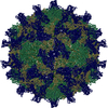
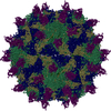
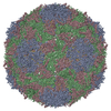
| ムービー |
 ムービービューア ムービービューア |
|---|---|
| 構造ビューア | 分子:  Molmil Molmil Jmol/JSmol Jmol/JSmol |
- ダウンロードとリンク
- ダウンロード
| PDBx/mmCIF形式 |  6gv4.cif.gz 6gv4.cif.gz | 306.9 KB | 表示 |  PDBx/mmCIF形式 PDBx/mmCIF形式 |
|---|---|---|---|---|
| PDB形式 |  pdb6gv4.ent.gz pdb6gv4.ent.gz | 242.7 KB | 表示 |  PDB形式 PDB形式 |
| PDBx/mmJSON形式 |  6gv4.json.gz 6gv4.json.gz | ツリー表示 |  PDBx/mmJSON形式 PDBx/mmJSON形式 | |
| その他 |  その他のダウンロード その他のダウンロード |
-検証レポート
| 文書・要旨 |  6gv4_validation.pdf.gz 6gv4_validation.pdf.gz | 1.1 MB | 表示 |  wwPDB検証レポート wwPDB検証レポート |
|---|---|---|---|---|
| 文書・詳細版 |  6gv4_full_validation.pdf.gz 6gv4_full_validation.pdf.gz | 1.1 MB | 表示 | |
| XML形式データ |  6gv4_validation.xml.gz 6gv4_validation.xml.gz | 31.2 KB | 表示 | |
| CIF形式データ |  6gv4_validation.cif.gz 6gv4_validation.cif.gz | 47.3 KB | 表示 | |
| アーカイブディレクトリ |  https://data.pdbj.org/pub/pdb/validation_reports/gv/6gv4 https://data.pdbj.org/pub/pdb/validation_reports/gv/6gv4 ftp://data.pdbj.org/pub/pdb/validation_reports/gv/6gv4 ftp://data.pdbj.org/pub/pdb/validation_reports/gv/6gv4 | HTTPS FTP |
-関連構造データ
| 関連構造データ |  0069MC M: このデータのモデリングに利用したマップデータ C: 同じ文献を引用 ( |
|---|---|
| 類似構造データ | |
| 電子顕微鏡画像生データ |  EMPIAR-10983 (タイトル: High-resolution Cryo-EM of Fab-labeled human parechovirus 3 EMPIAR-10983 (タイトル: High-resolution Cryo-EM of Fab-labeled human parechovirus 3Data size: 3.3 TB Data #1: Unaligned multi-frame micrographs of HPeV3-fab complex [micrographs - multiframe]) |
- リンク
リンク
- 集合体
集合体
| 登録構造単位 | 
|
|---|---|
| 1 | x 60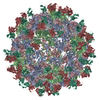
|
- 要素
要素
-RNA鎖 , 1種, 1分子 D
| #1: RNA鎖 | 分子量: 2505.489 Da / 分子数: 1 / 由来タイプ: 天然 / 詳細: RNA / 由来: (天然)  Human parechovirus 3 (ウイルス) / 細胞株: HT29 / 器官: colon adenocarcinoma Human parechovirus 3 (ウイルス) / 細胞株: HT29 / 器官: colon adenocarcinoma |
|---|
-タンパク質 , 3種, 3分子 ABC
| #2: タンパク質 | 分子量: 31770.135 Da / 分子数: 1 / 由来タイプ: 天然 / 由来: (天然)  Human parechovirus 3 (ウイルス) / 細胞株: HT29 / 器官: colon adenocarcinoma / 参照: UniProt: Q8BES5 Human parechovirus 3 (ウイルス) / 細胞株: HT29 / 器官: colon adenocarcinoma / 参照: UniProt: Q8BES5 |
|---|---|
| #3: タンパク質 | 分子量: 25913.127 Da / 分子数: 1 / 由来タイプ: 天然 / 由来: (天然)  Human parechovirus 3 (ウイルス) / 細胞株: HT29 / 器官: colon adenocarcinoma / 参照: UniProt: Q8BES5 Human parechovirus 3 (ウイルス) / 細胞株: HT29 / 器官: colon adenocarcinoma / 参照: UniProt: Q8BES5 |
| #4: タンパク質 | 分子量: 28757.551 Da / 分子数: 1 / 由来タイプ: 天然 / 詳細: polypeptide chain / 由来: (天然)  Human parechovirus 3 (ウイルス) / 細胞株: HT29 / 器官: colon adenocarcinoma / 参照: UniProt: Q8BES5, UniProt: A0A291FGQ4*PLUS Human parechovirus 3 (ウイルス) / 細胞株: HT29 / 器官: colon adenocarcinoma / 参照: UniProt: Q8BES5, UniProt: A0A291FGQ4*PLUS |
-抗体 , 2種, 2分子 HL
| #5: 抗体 | 分子量: 13128.613 Da / 分子数: 1 / 由来タイプ: 組換発現 / 詳細: polypeptide chain / 由来: (組換発現)  Homo sapiens (ヒト) / 細胞株 (発現宿主): 293T / 発現宿主: Homo sapiens (ヒト) / 細胞株 (発現宿主): 293T / 発現宿主:  Homo sapiens (ヒト) Homo sapiens (ヒト) |
|---|---|
| #6: 抗体 | 分子量: 12339.752 Da / 分子数: 1 / 由来タイプ: 組換発現 / 詳細: polypeptide chain / 由来: (組換発現)  Homo sapiens (ヒト) / 細胞株 (発現宿主): 293T / 発現宿主: Homo sapiens (ヒト) / 細胞株 (発現宿主): 293T / 発現宿主:  Homo sapiens (ヒト) Homo sapiens (ヒト) |
-詳細
| Has protein modification | Y |
|---|
-実験情報
-実験
| 実験 | 手法: 電子顕微鏡法 |
|---|---|
| EM実験 | 試料の集合状態: PARTICLE / 3次元再構成法: 単粒子再構成法 |
- 試料調製
試料調製
| 構成要素 |
| ||||||||||||||||||||||||
|---|---|---|---|---|---|---|---|---|---|---|---|---|---|---|---|---|---|---|---|---|---|---|---|---|---|
| 分子量 | 値: 7.7 MDa / 実験値: NO | ||||||||||||||||||||||||
| 由来(天然) |
| ||||||||||||||||||||||||
| 由来(組換発現) | 生物種:  Homo sapiens (ヒト) Homo sapiens (ヒト) | ||||||||||||||||||||||||
| ウイルスについての詳細 | 中空か: NO / エンベロープを持つか: NO / 単離: STRAIN / タイプ: VIRION | ||||||||||||||||||||||||
| 緩衝液 | pH: 7.5 | ||||||||||||||||||||||||
| 緩衝液成分 |
| ||||||||||||||||||||||||
| 試料 | 濃度: 0.1 mg/ml / 包埋: NO / シャドウイング: NO / 染色: NO / 凍結: YES | ||||||||||||||||||||||||
| 試料支持 | 詳細: ultrathin carbon-coated lacey 400-mesh copper grids (Ted Pella product #01824) グリッドの材料: COPPER / グリッドのサイズ: 400 divisions/in. | ||||||||||||||||||||||||
| 急速凍結 | 装置: HOMEMADE PLUNGER / 凍結剤: ETHANE / 凍結前の試料温度: 295 K 詳細: We could not control humidity during plunging. It was ambient humidity. Blot for 1 s before plunging. |
- 電子顕微鏡撮影
| 実験機器 |  モデル: Titan Krios / 画像提供: FEI Company |
|---|---|
| 顕微鏡 | モデル: FEI TITAN KRIOS |
| 電子銃 | 電子線源:  FIELD EMISSION GUN / 加速電圧: 300 kV / 照射モード: FLOOD BEAM FIELD EMISSION GUN / 加速電圧: 300 kV / 照射モード: FLOOD BEAM |
| 電子レンズ | モード: BRIGHT FIELD / 倍率(公称値): 75000 X / 最大 デフォーカス(公称値): 2500 nm / 最小 デフォーカス(公称値): 500 nm / Cs: 2.7 mm |
| 試料ホルダ | 凍結剤: NITROGEN 試料ホルダーモデル: FEI TITAN KRIOS AUTOGRID HOLDER |
| 撮影 | 平均露光時間: 1 sec. / 電子線照射量: 48 e/Å2 / 検出モード: INTEGRATING フィルム・検出器のモデル: FEI FALCON II (4k x 4k) 撮影したグリッド数: 2 / 実像数: 6541 |
| 画像スキャン | 動画フレーム数/画像: 18 / 利用したフレーム数/画像: 2-17 |
- 解析
解析
| EMソフトウェア |
| ||||||||||||||||||||||||||||||||||||||||||||||||||
|---|---|---|---|---|---|---|---|---|---|---|---|---|---|---|---|---|---|---|---|---|---|---|---|---|---|---|---|---|---|---|---|---|---|---|---|---|---|---|---|---|---|---|---|---|---|---|---|---|---|---|---|
| CTF補正 | 詳細: GCTF was used to estimate ctf / タイプ: PHASE FLIPPING AND AMPLITUDE CORRECTION | ||||||||||||||||||||||||||||||||||||||||||||||||||
| 粒子像の選択 | 選択した粒子像数: 217212 詳細: automatic particle selection in RELION using template generated from manually selected particles | ||||||||||||||||||||||||||||||||||||||||||||||||||
| 対称性 | 点対称性: I (正20面体型対称) | ||||||||||||||||||||||||||||||||||||||||||||||||||
| 3次元再構成 | 解像度: 2.8 Å / 解像度の算出法: FSC 0.143 CUT-OFF / 粒子像の数: 74927 / クラス平均像の数: 3 / 対称性のタイプ: POINT | ||||||||||||||||||||||||||||||||||||||||||||||||||
| 原子モデル構築 | プロトコル: OTHER / 空間: REAL 詳細: Initial model was generated in I-TASSER and SWISSMODEL using 4z92 and 4udf as reference. Initial rigid fit of the model to the map was done in UCSF Chimera. Model refinement was done in Coot and MDFF. |
 ムービー
ムービー コントローラー




 PDBj
PDBj


