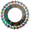
+ Open data
- Basic information
| Entry | Database: PDB / ID: 6dlw | ||||||
|---|---|---|---|---|---|---|---|
| Title | Complement component polyC9 | ||||||
 Components Components | Complement component C9 | ||||||
 Keywords Keywords | IMMUNE SYSTEM / Complement / pore / EM / membrane / transmembrane | ||||||
| Function / homology |  Function and homology information Function and homology informationcell killing / Terminal pathway of complement / membrane attack complex / other organism cell membrane / complement activation / complement activation, alternative pathway / complement activation, classical pathway / Regulation of Complement cascade / protein homooligomerization / positive regulation of immune response ...cell killing / Terminal pathway of complement / membrane attack complex / other organism cell membrane / complement activation / complement activation, alternative pathway / complement activation, classical pathway / Regulation of Complement cascade / protein homooligomerization / positive regulation of immune response / blood microparticle / killing of cells of another organism / extracellular space / extracellular exosome / extracellular region / plasma membrane Similarity search - Function | ||||||
| Biological species |  Homo sapiens (human) Homo sapiens (human) | ||||||
| Method | ELECTRON MICROSCOPY / single particle reconstruction / cryo EM / Resolution: 3.9 Å | ||||||
 Authors Authors | Dunstone, M.A. / Spicer, B.A. / Law, R.H.P. | ||||||
| Funding support |  Australia, 1items Australia, 1items
| ||||||
 Citation Citation |  Journal: Nat Commun / Year: 2018 Journal: Nat Commun / Year: 2018Title: The first transmembrane region of complement component-9 acts as a brake on its self-assembly. Authors: Bradley A Spicer / Ruby H P Law / Tom T Caradoc-Davies / Sue M Ekkel / Charles Bayly-Jones / Siew-Siew Pang / Paul J Conroy / Georg Ramm / Mazdak Radjainia / Hariprasad Venugopal / James C ...Authors: Bradley A Spicer / Ruby H P Law / Tom T Caradoc-Davies / Sue M Ekkel / Charles Bayly-Jones / Siew-Siew Pang / Paul J Conroy / Georg Ramm / Mazdak Radjainia / Hariprasad Venugopal / James C Whisstock / Michelle A Dunstone /    Abstract: Complement component 9 (C9) functions as the pore-forming component of the Membrane Attack Complex (MAC). During MAC assembly, multiple copies of C9 are sequentially recruited to membrane associated ...Complement component 9 (C9) functions as the pore-forming component of the Membrane Attack Complex (MAC). During MAC assembly, multiple copies of C9 are sequentially recruited to membrane associated C5b8 to form a pore. Here we determined the 2.2 Å crystal structure of monomeric murine C9 and the 3.9 Å resolution cryo EM structure of C9 in a polymeric assembly. Comparison with other MAC proteins reveals that the first transmembrane region (TMH1) in monomeric C9 is uniquely positioned and functions to inhibit its self-assembly in the absence of C5b8. We further show that following C9 recruitment to C5b8, a conformational change in TMH1 permits unidirectional and sequential binding of additional C9 monomers to the growing MAC. This mechanism of pore formation contrasts with related proteins, such as perforin and the cholesterol dependent cytolysins, where it is believed that pre-pore assembly occurs prior to the simultaneous release of the transmembrane regions. | ||||||
| History |
|
- Structure visualization
| Movie |
 Movie viewer Movie viewer |
|---|---|
| Structure viewer | Molecule:  Molmil Molmil Jmol/JSmol Jmol/JSmol |
- Downloads & links
- Download
| PDBx/mmCIF format |  6dlw.cif.gz 6dlw.cif.gz | 1.7 MB | Display |  PDBx/mmCIF format PDBx/mmCIF format |
|---|---|---|---|---|
| PDB format |  pdb6dlw.ent.gz pdb6dlw.ent.gz | 1.4 MB | Display |  PDB format PDB format |
| PDBx/mmJSON format |  6dlw.json.gz 6dlw.json.gz | Tree view |  PDBx/mmJSON format PDBx/mmJSON format | |
| Others |  Other downloads Other downloads |
-Validation report
| Arichive directory |  https://data.pdbj.org/pub/pdb/validation_reports/dl/6dlw https://data.pdbj.org/pub/pdb/validation_reports/dl/6dlw ftp://data.pdbj.org/pub/pdb/validation_reports/dl/6dlw ftp://data.pdbj.org/pub/pdb/validation_reports/dl/6dlw | HTTPS FTP |
|---|
-Related structure data
| Related structure data |  7773MC  6cxoC M: map data used to model this data C: citing same article ( |
|---|---|
| Similar structure data |
- Links
Links
- Assembly
| Deposited unit | 
|
|---|---|
| 1 |
|
- Components
| #1: Protein | Mass: 61056.594 Da / Num. of mol.: 22 Source method: isolated from a genetically manipulated source Source: (gene. exp.)  Homo sapiens (human) / Gene: C9 / Production host: Homo sapiens (human) / Gene: C9 / Production host:  Homo sapiens (human) / References: UniProt: P02748 Homo sapiens (human) / References: UniProt: P02748#2: Sugar | ChemComp-NAG / #3: Sugar | ChemComp-BMA / Has protein modification | Y | |
|---|
-Experimental details
-Experiment
| Experiment | Method: ELECTRON MICROSCOPY |
|---|---|
| EM experiment | Aggregation state: PARTICLE / 3D reconstruction method: single particle reconstruction |
- Sample preparation
| Component | Name: complement component polyC9 / Type: COMPLEX Details: PolyC9 component of the membrane attack complex (MAC) Entity ID: #1 / Source: RECOMBINANT | ||||||||||||
|---|---|---|---|---|---|---|---|---|---|---|---|---|---|
| Molecular weight | Value: 1100 kDa/nm / Experimental value: NO | ||||||||||||
| Source (natural) | Organism:  Homo sapiens (human) Homo sapiens (human) | ||||||||||||
| Source (recombinant) | Organism:  Homo sapiens (human) Homo sapiens (human) | ||||||||||||
| Buffer solution | pH: 7.5 | ||||||||||||
| Buffer component |
| ||||||||||||
| Specimen | Conc.: 1.3 mg/ml / Embedding applied: NO / Shadowing applied: NO / Staining applied: NO / Vitrification applied: YES / Details: The sample was monodisperse | ||||||||||||
| Specimen support | Details: Negative glow discharge / Grid material: COPPER / Grid mesh size: 200 divisions/in. / Grid type: Quantifoil R1.2/1.3 | ||||||||||||
| Vitrification | Instrument: FEI VITROBOT MARK IV / Cryogen name: ETHANE / Humidity: 100 % / Chamber temperature: 277.15 K / Details: blot for 2 seconds before plunging |
- Electron microscopy imaging
| Experimental equipment |  Model: Titan Krios / Image courtesy: FEI Company |
|---|---|
| Microscopy | Model: FEI TITAN KRIOS |
| Electron gun | Electron source:  FIELD EMISSION GUN / Accelerating voltage: 300 kV / Illumination mode: FLOOD BEAM FIELD EMISSION GUN / Accelerating voltage: 300 kV / Illumination mode: FLOOD BEAM |
| Electron lens | Mode: BRIGHT FIELD |
| Image recording | Average exposure time: 8 sec. / Electron dose: 46.4 e/Å2 / Detector mode: SUPER-RESOLUTION / Film or detector model: GATAN K2 SUMMIT (4k x 4k) |
- Processing
| EM software |
| ||||||||||||||||||||||||
|---|---|---|---|---|---|---|---|---|---|---|---|---|---|---|---|---|---|---|---|---|---|---|---|---|---|
| CTF correction | Type: PHASE FLIPPING AND AMPLITUDE CORRECTION | ||||||||||||||||||||||||
| Symmetry | Point symmetry: C22 (22 fold cyclic) | ||||||||||||||||||||||||
| 3D reconstruction | Resolution: 3.9 Å / Resolution method: FSC 0.143 CUT-OFF / Num. of particles: 58000 / Symmetry type: POINT | ||||||||||||||||||||||||
| Atomic model building | Protocol: RIGID BODY FIT |
 Movie
Movie Controller


 PDBj
PDBj


