 Movie
Movie Controller

[English] 日本語
 Yorodumi
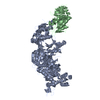
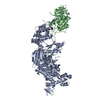
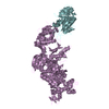
- PDB-6c0v: Molecular structure of human P-glycoprotein in the ATP-bound, out... -
+ Open data

- Basic information
| Entry | Database: PDB / ID: 6c0v | ||||||
|---|---|---|---|---|---|---|---|
| Title | Molecular structure of human P-glycoprotein in the ATP-bound, outward-facing conformation | ||||||
 Components Components | Multidrug resistance protein 1 | ||||||
 Keywords Keywords | TRANSPORT PROTEIN / ABC transporter / ABCB1 / P-glycoprotein / cryo-EM / multidrug resistance / Structural Genomics / PSI-2 / Protein Structure Initiative | ||||||
| Function / homology |  Function and homology information Function and homology informationhormone transport / phosphatidylethanolamine floppase activity / cellular response to nonylphenol / cellular response to borneol / response to codeine / cellular response to mycotoxin / daunorubicin transport / positive regulation of response to drug / terpenoid transport / ceramide floppase activity ...hormone transport / phosphatidylethanolamine floppase activity / cellular response to nonylphenol / cellular response to borneol / response to codeine / cellular response to mycotoxin / daunorubicin transport / positive regulation of response to drug / terpenoid transport / ceramide floppase activity / regulation of intestinal absorption / cellular response to external biotic stimulus / response to cyclosporin A / response to antineoplastic agent / positive regulation of establishment of Sertoli cell barrier / negative regulation of sensory perception of pain / carboxylic acid transmembrane transport / floppase activity / ceramide translocation / Abacavir transmembrane transport / response to quercetin / carboxylic acid transmembrane transporter activity / establishment of blood-retinal barrier / phosphatidylethanolamine flippase activity / protein localization to bicellular tight junction / phosphatidylcholine floppase activity / external side of apical plasma membrane / Atorvastatin ADME / response to thyroxine / xenobiotic transport across blood-brain barrier / establishment of blood-brain barrier / export across plasma membrane / P-type phospholipid transporter / xenobiotic detoxification by transmembrane export across the plasma membrane / transepithelial transport / cellular response to L-glutamate / response to vitamin A / ABC-type xenobiotic transporter / response to glucagon / response to vitamin D / intestinal absorption / response to alcohol / response to glycoside / Prednisone ADME / phospholipid translocation / ABC-type xenobiotic transporter activity / cellular hyperosmotic salinity response / cellular response to alkaloid / cellular response to antibiotic / maintenance of blood-brain barrier / efflux transmembrane transporter activity / ATPase-coupled transmembrane transporter activity / xenobiotic transmembrane transporter activity / cellular response to dexamethasone stimulus / response to cadmium ion / transmembrane transporter activity / transport across blood-brain barrier / lactation / response to progesterone / xenobiotic metabolic process / regulation of chloride transport / placenta development / stem cell proliferation / cellular response to estradiol stimulus / brush border membrane / female pregnancy / circadian rhythm / transmembrane transport / G2/M transition of mitotic cell cycle / cellular response to tumor necrosis factor / ABC-family protein mediated transport / cellular response to lipopolysaccharide / response to hypoxia / apical plasma membrane / response to xenobiotic stimulus / ubiquitin protein ligase binding / cell surface / ATP hydrolysis activity / extracellular exosome / ATP binding / membrane / plasma membrane / cytoplasm Similarity search - Function | ||||||
| Biological species |  Homo sapiens (human) Homo sapiens (human) | ||||||
| Method | ELECTRON MICROSCOPY / single particle reconstruction / cryo EM / Resolution: 3.4 Å | ||||||
 Authors Authors | Kim, Y.J. / Chen, J. | ||||||
| Funding support |  United States, 1items United States, 1items
| ||||||
 Citation Citation | Journal: Science / Year: 2018 Title: Molecular structure of human P-glycoprotein in the ATP-bound, outward-facing conformation. Authors: Youngjin Kim / Jue Chen / Abstract: The multidrug transporter permeability (P)-glycoprotein is an adenosine triphosphate (ATP)-binding cassette exporter responsible for clinical resistance to chemotherapy. P-glycoprotein extrudes toxic ...The multidrug transporter permeability (P)-glycoprotein is an adenosine triphosphate (ATP)-binding cassette exporter responsible for clinical resistance to chemotherapy. P-glycoprotein extrudes toxic molecules and drugs from cells through ATP-powered conformational changes. Despite decades of effort, only the structures of the inward-facing conformation of P-glycoprotein are available. Here we present the structure of human P-glycoprotein in the outward-facing conformation, determined by cryo-electron microscopy at 3.4-angstrom resolution. The two nucleotide-binding domains form a closed dimer occluding two ATP molecules. The drug-binding cavity observed in the inward-facing structures is reorientated toward the extracellular space and compressed to preclude substrate binding. This observation indicates that ATP binding, not hydrolysis, promotes substrate release. The structure evokes a model in which the dynamic nature of P-glycoprotein enables translocation of a large variety of substrates. | ||||||
| History |
|
- Structure visualization
| Movie |
Movie viewer |
|---|---|
| Structure viewer | Molecule: MolmilJmol/JSmol |
- Downloads & links
-Download
| PDBx/mmCIF format | 6c0v.cif.gz | 241.9 KB | Display | PDBx/mmCIF format |
|---|---|---|---|---|
| PDB format | pdb6c0v.ent.gz | 186.5 KB | Display | PDB format |
| PDBx/mmJSON format | 6c0v.json.gz | Tree view | PDBx/mmJSON format | |
| Others |  Other downloads Other downloads |
-Validation report
| Arichive directory | https://data.pdbj.org/pub/pdb/validation_reports/c0/6c0vftp://data.pdbj.org/pub/pdb/validation_reports/c0/6c0v | HTTPS FTP |
|---|
-Related structure data
| Related structure data |  7325MC M: map data used to model this data C: citing same article ( |
|---|---|
| Similar structure data | |
| EM raw data | EMPIAR-10803 (Title: Cryo-electron microscopy reconstruction of ATP-bound human P-glycoprotein Data size: 2.0 TB Data #1: Unaligned and uncorrected multiframe movies of human ATP-bound P-glycoprotein [micrographs - multiframe]) |


-Links
 PDBj
PDBj

- Assembly
| Deposited unit | 
|
|---|---|
| 1 |
|
-Components
| #1: Protein | Mass: 142660.922 Da / Num. of mol.: 1 / Mutation: E556Q, E1201Q Source method: isolated from a genetically manipulated source Source: (gene. exp.) Homo sapiens (human) / Gene: ABCB1, MDR1, PGY1 / Cell line (production host): HEK293S GnTI- / Production host: Homo sapiens (human) / References: UniProt: P08183, EC: 3.6.3.44 | ||
|---|---|---|---|
| #2: Chemical |   Mass: 507.181 Da / Num. of mol.: 2 / Source method: obtained synthetically / Formula: C10H16N5O13P3 / Comment: ATP, energy-carrying molecule*YM Mass: 507.181 Da / Num. of mol.: 2 / Source method: obtained synthetically / Formula: C10H16N5O13P3 / Comment: ATP, energy-carrying molecule*YM#3: Chemical |   Mass: 24.305 Da / Num. of mol.: 2 / Source method: obtained synthetically / Formula: Mg Mass: 24.305 Da / Num. of mol.: 2 / Source method: obtained synthetically / Formula: Mg |
-Experimental details
-Experiment
| Experiment | Method: ELECTRON MICROSCOPY |
|---|---|
| EM experiment | Aggregation state: PARTICLE / 3D reconstruction method: single particle reconstruction |
- Sample preparation
| Component | Name: Human P-glycoprotein E556Q, E1201Q / Type: COMPLEX / Entity ID: #1 / Source: RECOMBINANT | ||||||||||||||||||||||||||||||||||||
|---|---|---|---|---|---|---|---|---|---|---|---|---|---|---|---|---|---|---|---|---|---|---|---|---|---|---|---|---|---|---|---|---|---|---|---|---|---|
| Molecular weight | Value: 0.141 MDa / Experimental value: YES | ||||||||||||||||||||||||||||||||||||
| Source (natural) | Organism: Homo sapiens (human) | ||||||||||||||||||||||||||||||||||||
| Source (recombinant) | Organism: Homo sapiens (human) | ||||||||||||||||||||||||||||||||||||
| Buffer solution | pH: 7.5 | ||||||||||||||||||||||||||||||||||||
| Buffer component |
| ||||||||||||||||||||||||||||||||||||
| Specimen | Conc.: 5 mg/ml / Embedding applied: NO / Shadowing applied: NO / Staining applied: NO / Vitrification applied: YES | ||||||||||||||||||||||||||||||||||||
| Specimen support | Grid material: GOLD / Grid type: Quantifoil R1.2/1.3 | ||||||||||||||||||||||||||||||||||||
| Vitrification | Instrument: FEI VITROBOT MARK IV / Cryogen name: ETHANE / Humidity: 100 % / Chamber temperature: 295 K |
- Electron microscopy imaging
| Experimental equipment |  Model: Titan Krios / Image courtesy: FEI Company |
|---|---|
| Microscopy | Model: FEI TITAN KRIOS |
| Electron gun | Electron source:  FIELD EMISSION GUN / Accelerating voltage: 300 kV / Illumination mode: FLOOD BEAM FIELD EMISSION GUN / Accelerating voltage: 300 kV / Illumination mode: FLOOD BEAM |
| Electron lens | Mode: BRIGHT FIELD / Nominal defocus max: 2200 nm / Nominal defocus min: 800 nm / Cs: 2.7 mm / C2 aperture diameter: 70 µm |
| Specimen holder | Cryogen: NITROGEN / Specimen holder model: FEI TITAN KRIOS AUTOGRID HOLDER / Temperature (max): 100 K / Temperature (min): 80 K |
| Image recording | Average exposure time: 7 sec. / Electron dose: 80 e/Å2 / Detector mode: SUPER-RESOLUTION / Film or detector model: GATAN K2 SUMMIT (4k x 4k) / Num. of grids imaged: 2 / Num. of real images: 5747 |
| EM imaging optics | Energyfilter upper: 10 eV / Energyfilter lower: 10 eV |
| Image scans | Width: 3838 / Height: 3710 / Movie frames/image: 50 / Used frames/image: 1-50 |
- Processing
| Software | Name: REFMAC / Version: 5.8.0189 / Classification: refinement | ||||||||||||||||||||||||||||||||||||||||||||||||||||||||||||||||||||||||||||||||||||||||||||||||||||||||||
|---|---|---|---|---|---|---|---|---|---|---|---|---|---|---|---|---|---|---|---|---|---|---|---|---|---|---|---|---|---|---|---|---|---|---|---|---|---|---|---|---|---|---|---|---|---|---|---|---|---|---|---|---|---|---|---|---|---|---|---|---|---|---|---|---|---|---|---|---|---|---|---|---|---|---|---|---|---|---|---|---|---|---|---|---|---|---|---|---|---|---|---|---|---|---|---|---|---|---|---|---|---|---|---|---|---|---|---|
| EM software |
| ||||||||||||||||||||||||||||||||||||||||||||||||||||||||||||||||||||||||||||||||||||||||||||||||||||||||||
| CTF correction | Type: PHASE FLIPPING AND AMPLITUDE CORRECTION | ||||||||||||||||||||||||||||||||||||||||||||||||||||||||||||||||||||||||||||||||||||||||||||||||||||||||||
| Particle selection | Num. of particles selected: 143451 | ||||||||||||||||||||||||||||||||||||||||||||||||||||||||||||||||||||||||||||||||||||||||||||||||||||||||||
| Symmetry | Point symmetry: C1 (asymmetric) | ||||||||||||||||||||||||||||||||||||||||||||||||||||||||||||||||||||||||||||||||||||||||||||||||||||||||||
| 3D reconstruction | Resolution: 3.4 Å / Resolution method: FSC 0.143 CUT-OFF / Num. of particles: 143451 / Symmetry type: POINT | ||||||||||||||||||||||||||||||||||||||||||||||||||||||||||||||||||||||||||||||||||||||||||||||||||||||||||
| Atomic model building | Protocol: FLEXIBLE FIT / Space: RECIPROCAL | ||||||||||||||||||||||||||||||||||||||||||||||||||||||||||||||||||||||||||||||||||||||||||||||||||||||||||
| Refinement | Resolution: 3.4→145.61 Å / Cor.coef. Fo:Fc: 0.979 / ESU R: 0.47 Stereochemistry target values: MAXIMUM LIKELIHOOD WITH PHASES Details: HYDROGENS HAVE BEEN ADDED IN THE RIDING POSITIONS
| ||||||||||||||||||||||||||||||||||||||||||||||||||||||||||||||||||||||||||||||||||||||||||||||||||||||||||
| Solvent computation | Ion probe radii: 0.8 Å / Shrinkage radii: 0.8 Å / VDW probe radii: 1.2 Å / Solvent model: MASK | ||||||||||||||||||||||||||||||||||||||||||||||||||||||||||||||||||||||||||||||||||||||||||||||||||||||||||
| Displacement parameters | Biso mean: 266.758 Å2
| ||||||||||||||||||||||||||||||||||||||||||||||||||||||||||||||||||||||||||||||||||||||||||||||||||||||||||
| Refinement step | Cycle: 1 / Total: 8976 | ||||||||||||||||||||||||||||||||||||||||||||||||||||||||||||||||||||||||||||||||||||||||||||||||||||||||||
| Refine LS restraints |
|