 Movie
Movie Controller

[English] 日本語
 Yorodumi
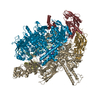
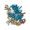
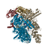
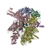
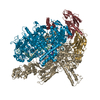
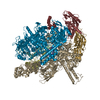
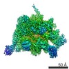
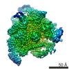
- PDB-6alg: CryoEM structure of HK022 Nun - E.coli RNA polymerase elongation ... -
+ Open data

- Basic information
| Entry | Database: PDB / ID: 6alg | |||||||||
|---|---|---|---|---|---|---|---|---|---|---|
| Title | CryoEM structure of HK022 Nun - E.coli RNA polymerase elongation complex | |||||||||
 Components Components |
| |||||||||
 Keywords Keywords | transcription/dna/rna / DNA-dependent RNA polymerase / TRANSCRIPTION / transcription-dna-rna complex | |||||||||
| Function / homology |  Function and homology information Function and homology informationnegative regulation of DNA-templated transcription, elongation / RNA polymerase complex / submerged biofilm formation / cellular response to cell envelope stress / regulation of DNA-templated transcription initiation / bacterial-type flagellum assembly / bacterial-type RNA polymerase core enzyme binding / cytosolic DNA-directed RNA polymerase complex / bacterial-type flagellum-dependent cell motility / nitrate assimilation ...negative regulation of DNA-templated transcription, elongation / RNA polymerase complex / submerged biofilm formation / cellular response to cell envelope stress / regulation of DNA-templated transcription initiation / bacterial-type flagellum assembly / bacterial-type RNA polymerase core enzyme binding / cytosolic DNA-directed RNA polymerase complex / bacterial-type flagellum-dependent cell motility / nitrate assimilation / regulation of DNA-templated transcription elongation / transcription elongation factor complex / DNA-directed RNA polymerase complex / transcription antitermination / cell motility / DNA-templated transcription initiation / DNA-templated transcription termination / ribonucleoside binding / DNA-directed RNA polymerase / DNA-directed RNA polymerase activity / response to heat / protein-containing complex assembly / intracellular iron ion homeostasis / protein dimerization activity / response to antibiotic / DNA-templated transcription / magnesium ion binding / DNA binding / zinc ion binding / membrane / cytoplasm / cytosol Similarity search - Function | |||||||||
| Biological species |   Escherichia phage HK022 (virus) Escherichia phage HK022 (virus) Enterobacteria phage T7 (virus) Enterobacteria phage T7 (virus) | |||||||||
| Method | ELECTRON MICROSCOPY / single particle reconstruction / cryo EM / Resolution: 3.7 Å | |||||||||
 Authors Authors | Kang, J.Y. / Darst, S.A. | |||||||||
| Funding support |  United States, 1items United States, 1items
| |||||||||
 Citation Citation | Journal: Elife / Year: 2017 Title: Structural basis of transcription arrest by coliphage HK022 Nun in an RNA polymerase elongation complex. Authors: Jin Young Kang / Paul Dominic B Olinares / James Chen / Elizabeth A Campbell / Arkady Mustaev / Brian T Chait / Max E Gottesman / Seth A Darst / Abstract: Coliphage HK022 Nun blocks superinfection by coliphage λ by stalling RNA polymerase (RNAP) translocation specifically on λ DNA. To provide a structural framework to understand how Nun blocks RNAP ...Coliphage HK022 Nun blocks superinfection by coliphage λ by stalling RNA polymerase (RNAP) translocation specifically on λ DNA. To provide a structural framework to understand how Nun blocks RNAP translocation, we determined structures of RNAP ternary elongation complexes (TECs) with and without Nun by single-particle cryo-electron microscopy. Nun fits tightly into the TEC by taking advantage of gaps between the RNAP and the nucleic acids. The C-terminal segment of Nun interacts with the RNAP β and β' subunits inside the RNAP active site cleft as well as with nearly every element of the nucleic acid scaffold, essentially crosslinking the RNAP and the nucleic acids to prevent translocation, a mechanism supported by the effects of Nun amino acid substitutions. The nature of Nun interactions inside the RNAP active site cleft suggests that RNAP clamp opening is required for Nun to establish its interactions, explaining why Nun acts on paused TECs. | |||||||||
| History |
|
- Structure visualization
| Movie |
Movie viewer |
|---|---|
| Structure viewer | Molecule: MolmilJmol/JSmol |
- Downloads & links
-Download
| PDBx/mmCIF format | 6alg.cif.gz | 600.2 KB | Display | PDBx/mmCIF format |
|---|---|---|---|---|
| PDB format | pdb6alg.ent.gz | 475.6 KB | Display | PDB format |
| PDBx/mmJSON format | 6alg.json.gz | Tree view | PDBx/mmJSON format | |
| Others |  Other downloads Other downloads |
-Validation report
| Arichive directory | https://data.pdbj.org/pub/pdb/validation_reports/al/6algftp://data.pdbj.org/pub/pdb/validation_reports/al/6alg | HTTPS FTP |
|---|
-Related structure data
| Related structure data |  8584MC 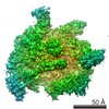 8585C 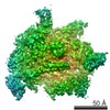 8586C 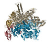 6alfC 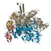 6alhC M: map data used to model this data C: citing same article ( |
|---|---|
| Similar structure data |


-Links
 PDBj
PDBj


- Assembly
| Deposited unit | 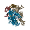
|
|---|---|
| 1 |
|
-Components
-DNA chain , 2 types, 2 molecules AB
| #1: DNA chain | Mass: 8840.689 Da / Num. of mol.: 1 / Source method: obtained synthetically / Source: (synth.) Enterobacteria phage T7 (virus) |
|---|---|
| #2: DNA chain | Mass: 8813.646 Da / Num. of mol.: 1 / Source method: obtained synthetically / Source: (synth.) Enterobacteria phage T7 (virus) |
-DNA-directed RNA polymerase subunit ... , 4 types, 5 molecules GHIJK
| #4: Protein | Mass: 26459.125 Da / Num. of mol.: 2 Source method: isolated from a genetically manipulated source Source: (gene. exp.) Strain: K12 / Gene: rpoA, pez, phs, sez, b3295, JW3257 / Production host: #5: Protein | | Mass: 150820.875 Da / Num. of mol.: 1 Source method: isolated from a genetically manipulated source Source: (gene. exp.) Strain: K12 Gene: rpoB, groN, nitB, rif, ron, stl, stv, tabD, b3987, JW3950 Production host: #6: Protein | | Mass: 155366.781 Da / Num. of mol.: 1 Source method: isolated from a genetically manipulated source Source: (gene. exp.) Strain: K12 / Gene: rpoC, tabB, b3988, JW3951 / Production host: #7: Protein | | Mass: 10249.547 Da / Num. of mol.: 1 Source method: isolated from a genetically manipulated source Source: (gene. exp.) Strain: K12 / Gene: rpoZ, b3649, JW3624 / Production host: |
|---|
-RNA chain / Protein , 2 types, 2 molecules RN
| #3: RNA chain | Mass: 6509.968 Da / Num. of mol.: 1 / Source method: obtained synthetically / Source: (synth.) Enterobacteria phage T7 (virus) |
|---|---|
| #8: Protein | Mass: 12758.371 Da / Num. of mol.: 1 Source method: isolated from a genetically manipulated source Source: (gene. exp.) Escherichia phage HK022 (virus) / Gene: nun / Production host: |
-Non-polymers , 2 types, 3 molecules 

| #9: Chemical | ChemComp-MG /  Mass: 24.305 Da / Num. of mol.: 1 / Source method: obtained synthetically / Formula: Mg Mass: 24.305 Da / Num. of mol.: 1 / Source method: obtained synthetically / Formula: Mg |
|---|---|
| #10: Chemical |  Mass: 65.409 Da / Num. of mol.: 2 / Source method: obtained synthetically / Formula: Zn Mass: 65.409 Da / Num. of mol.: 2 / Source method: obtained synthetically / Formula: Zn |
-Experimental details
-Experiment
| Experiment | Method: ELECTRON MICROSCOPY |
|---|---|
| EM experiment | Aggregation state: PARTICLE / 3D reconstruction method: single particle reconstruction |
- Sample preparation
| Component | Name: HK022 Nun - E.coli RNA polymerase elongation complex / Type: COMPLEX / Entity ID: #1-#8 / Source: MULTIPLE SOURCES |
|---|---|
| Molecular weight | Value: 0.406 MDa / Experimental value: NO |
| Source (natural) | Organism: |
| Source (recombinant) | Organism: |
| Buffer solution | pH: 8 Details: 20 mM Tris pH8.0, 150 mM Potassium Chloride, 5 mM Magnesium Chloride, 5 mM Dithreitol |
| Specimen | Conc.: 3 mg/ml / Embedding applied: NO / Shadowing applied: NO / Staining applied: NO / Vitrification applied: YES |
| Specimen support | Grid material: GOLD / Grid mesh size: 400 divisions/in. / Grid type: C-flat CF1.2/1.3 |
| Vitrification | Instrument: FEI VITROBOT MARK IV / Cryogen name: ETHANE / Humidity: 100 % / Chamber temperature: 295.15 K |
- Electron microscopy imaging
| Experimental equipment |  Model: Titan Krios / Image courtesy: FEI Company |
|---|---|
| Microscopy | Model: FEI TITAN KRIOS |
| Electron gun | Electron source:  FIELD EMISSION GUN / Accelerating voltage: 300 kV / Illumination mode: FLOOD BEAM FIELD EMISSION GUN / Accelerating voltage: 300 kV / Illumination mode: FLOOD BEAM |
| Electron lens | Mode: BRIGHT FIELD |
| Image recording | Electron dose: 89 e/Å2 / Detector mode: SUPER-RESOLUTION / Film or detector model: GATAN K2 SUMMIT (4k x 4k) |
| Image scans | Width: 3710 / Height: 3838 |
- Processing
| Software | Name: PHENIX / Version: 1.11.1_2575: / Classification: refinement | ||||||||||||||||||||||||
|---|---|---|---|---|---|---|---|---|---|---|---|---|---|---|---|---|---|---|---|---|---|---|---|---|---|
| EM software |
| ||||||||||||||||||||||||
| CTF correction | Details: Used CTFFIND4 / Type: NONE | ||||||||||||||||||||||||
| Symmetry | Point symmetry: C1 (asymmetric) | ||||||||||||||||||||||||
| 3D reconstruction | Resolution: 3.7 Å / Resolution method: FSC 0.143 CUT-OFF / Num. of particles: 192000 / Symmetry type: POINT | ||||||||||||||||||||||||
| Atomic model building | Protocol: RIGID BODY FIT | ||||||||||||||||||||||||
| Refine LS restraints |
|
