 Movie
Movie Controller


+ Open data

- Basic information
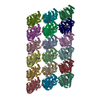
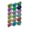
| Entry | Database: PDB / ID: 5xxx | |||||||||||||||
|---|---|---|---|---|---|---|---|---|---|---|---|---|---|---|---|---|
| Title | GMPCPP-microtubule complexed with nucleotide-free KIF5C | |||||||||||||||
 Components Components |
| |||||||||||||||
 Keywords Keywords | STRUCTURAL PROTEIN / Microtubule / KIF5C / Kinesin | |||||||||||||||
| Function / homology |  Function and homology information Function and homology informationstructural constituent of cytoskeleton / microtubule cytoskeleton organization / neuron migration / mitotic cell cycle / microtubule / Hydrolases; Acting on acid anhydrides; Acting on GTP to facilitate cellular and subcellular movement / hydrolase activity / GTPase activity / GTP binding / metal ion binding / cytoplasm Similarity search - Function | |||||||||||||||
| Biological species |  | |||||||||||||||
| Method | ELECTRON MICROSCOPY / helical reconstruction / cryo EM / Resolution: 6.43 Å | |||||||||||||||
 Authors Authors | Morikawa, M. / Shigematsu, H. / Nitta, R. / Hirokawa, N. | |||||||||||||||
| Funding support |  Japan, 4items Japan, 4items
| |||||||||||||||
 Citation Citation | Journal: J Cell Biol / Year: 2018 Title: Kinesin-binding-triggered conformation switching of microtubules contributes to polarized transport. Authors: Tomohiro Shima / Manatsu Morikawa / Junichi Kaneshiro / Taketoshi Kambara / Shinji Kamimura / Toshiki Yagi / Hiroyuki Iwamoto / Sotaro Uemura / Hideki Shigematsu / Mikako Shirouzu / Taro ...Authors: Tomohiro Shima / Manatsu Morikawa / Junichi Kaneshiro / Taketoshi Kambara / Shinji Kamimura / Toshiki Yagi / Hiroyuki Iwamoto / Sotaro Uemura / Hideki Shigematsu / Mikako Shirouzu / Taro Ichimura / Tomonobu M Watanabe / Ryo Nitta / Yasushi Okada / Nobutaka Hirokawa /  Abstract: Kinesin-1, the founding member of the kinesin superfamily of proteins, is known to use only a subset of microtubules for transport in living cells. This biased use of microtubules is proposed as the ...Kinesin-1, the founding member of the kinesin superfamily of proteins, is known to use only a subset of microtubules for transport in living cells. This biased use of microtubules is proposed as the guidance cue for polarized transport in neurons, but the underlying mechanisms are still poorly understood. Here, we report that kinesin-1 binding changes the microtubule lattice and promotes further kinesin-1 binding. This high-affinity state requires the binding of kinesin-1 in the nucleotide-free state. Microtubules return to the initial low-affinity state by washing out the binding kinesin-1 or by the binding of non-hydrolyzable ATP analogue AMPPNP to kinesin-1. X-ray fiber diffraction, fluorescence speckle microscopy, and second-harmonic generation microscopy, as well as cryo-EM, collectively demonstrated that the binding of nucleotide-free kinesin-1 to GDP microtubules changes the conformation of the GDP microtubule to a conformation resembling the GTP microtubule. | |||||||||||||||
| History |
|
- Structure visualization
| Movie |
Movie viewer |
|---|---|
| Structure viewer | Molecule: MolmilJmol/JSmol |
- Downloads & links
-Download
| PDBx/mmCIF format | 5xxx.cif.gz | 1.3 MB | Display | PDBx/mmCIF format |
|---|---|---|---|---|
| PDB format | pdb5xxx.ent.gz | 1 MB | Display | PDB format |
| PDBx/mmJSON format | 5xxx.json.gz | Tree view | PDBx/mmJSON format | |
| Others |  Other downloads Other downloads |
-Validation report
| Arichive directory | https://data.pdbj.org/pub/pdb/validation_reports/xx/5xxxftp://data.pdbj.org/pub/pdb/validation_reports/xx/5xxx | HTTPS FTP |
|---|
-Related structure data
| Related structure data | 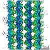 6783MC 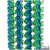 6779C 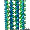 6781C 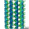 6782C 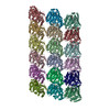 5xxtC 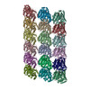 5xxvC  5xxwC C: citing same article ( M: map data used to model this data |
|---|---|
| Similar structure data |

-Links
 PDBj
PDBj


- Assembly
| Deposited unit | 
|
|---|---|
| 1 |
|
-Components
-Protein , 2 types, 18 molecules ACEGIKMOQBDFHJLNPR
| #1: Protein | Mass: 48638.793 Da / Num. of mol.: 9 / Fragment: UNP residues 2-439 / Source method: isolated from a natural source / Source: (natural) #2: Protein | Mass: 47809.746 Da / Num. of mol.: 9 / Fragment: UNP residues 2-427 / Source method: isolated from a natural source / Source: (natural) |
|---|
-Non-polymers , 4 types, 72 molecules 


| #3: Chemical | ChemComp-GTP /  Mass: 523.180 Da / Num. of mol.: 9 / Source method: obtained synthetically / Formula: C10H16N5O14P3 / Comment: GTP, energy-carrying molecule*YM Mass: 523.180 Da / Num. of mol.: 9 / Source method: obtained synthetically / Formula: C10H16N5O14P3 / Comment: GTP, energy-carrying molecule*YM#4: Chemical | ChemComp-MG /  Mass: 24.305 Da / Num. of mol.: 18 / Source method: obtained synthetically / Formula: Mg Mass: 24.305 Da / Num. of mol.: 18 / Source method: obtained synthetically / Formula: Mg#5: Chemical | ChemComp-G2P /  Mass: 521.208 Da / Num. of mol.: 9 / Source method: obtained synthetically / Formula: C11H18N5O13P3 / Comment: GMP-CPP, energy-carrying molecule analogue*YM Mass: 521.208 Da / Num. of mol.: 9 / Source method: obtained synthetically / Formula: C11H18N5O13P3 / Comment: GMP-CPP, energy-carrying molecule analogue*YM#6: Water | ChemComp-HOH / | Mass: 18.015 Da / Num. of mol.: 36 / Source method: isolated from a natural source / Formula: H2O |
|---|
-Details
| Has protein modification | Y |
|---|
-Experimental details
-Experiment
| Experiment | Method: ELECTRON MICROSCOPY |
|---|---|
| EM experiment | Aggregation state: FILAMENT / 3D reconstruction method: helical reconstruction |
- Sample preparation
| Component | Name: GMPCPP-microtubule complexed with nucleotide-free KIF5C Type: ORGANELLE OR CELLULAR COMPONENT / Entity ID: #1-#2 / Source: MULTIPLE SOURCES |
|---|---|
| Molecular weight | Experimental value: NO |
| Source (natural) | Organism: |
| Source (recombinant) | Organism: Plasmid: pET21b |
| Buffer solution | pH: 7.4 |
| Specimen | Embedding applied: NO / Shadowing applied: NO / Staining applied: NO / Vitrification applied: YES |
| Specimen support | Grid material: COPPER / Grid mesh size: 200 divisions/in. / Grid type: Quantifoil R2/2 |
| Vitrification | Instrument: FEI VITROBOT MARK IV / Cryogen name: ETHANE / Humidity: 100 % / Chamber temperature: 300 K |
- Electron microscopy imaging
| Experimental equipment |  Model: Talos Arctica / Image courtesy: FEI Company |
|---|---|
| Microscopy | Model: FEI TECNAI ARCTICA |
| Electron gun | Electron source:  FIELD EMISSION GUN / Accelerating voltage: 200 kV / Illumination mode: FLOOD BEAM FIELD EMISSION GUN / Accelerating voltage: 200 kV / Illumination mode: FLOOD BEAM |
| Electron lens | Mode: BRIGHT FIELD / Cs: 2.7 mm |
| Image recording | Electron dose: 50 e/Å2 / Film or detector model: FEI FALCON II (4k x 4k) |
- Processing
| EM software | Name: FREALIGN / Version: 9 / Category: 3D reconstruction |
|---|---|
| CTF correction | Type: PHASE FLIPPING AND AMPLITUDE CORRECTION |
| Helical symmerty | Angular rotation/subunit: -25.7541 ° / Axial rise/subunit: 8.8632 Å / Axial symmetry: C1 |
| 3D reconstruction | Resolution: 6.43 Å / Resolution method: FSC 0.143 CUT-OFF / Num. of particles: 5822 / Symmetry type: HELICAL |
| Atomic model building | Protocol: RIGID BODY FIT |
