National Institutes of Health/National Institute of General Medical Sciences (NIH/NIGMS)
P41GM103832
米国
National Institutes of Health/National Institute of General Medical Sciences (NIH/NIGMS)
U24GM116787
米国
Public Health Service
GM27099
米国
引用
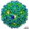
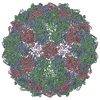
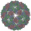
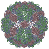
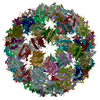
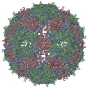
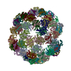
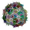
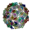
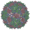
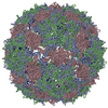
ジャーナル: Proc Natl Acad Sci U S A / 年: 2017 タイトル: Structures of Qβ virions, virus-like particles, and the Qβ-MurA complex reveal internal coat proteins and the mechanism of host lysis. 著者: Zhicheng Cui / Karl V Gorzelnik / Jeng-Yih Chang / Carrie Langlais / Joanita Jakana / Ry Young / Junjie Zhang / 要旨: In single-stranded RNA bacteriophages (ssRNA phages) a single copy of the maturation protein binds the genomic RNA (gRNA) and is required for attachment of the phage to the host pilus. For the ...In single-stranded RNA bacteriophages (ssRNA phages) a single copy of the maturation protein binds the genomic RNA (gRNA) and is required for attachment of the phage to the host pilus. For the canonical Qβ the maturation protein, A, has an additional role as the lysis protein, by its ability to bind and inhibit MurA, which is involved in peptidoglycan biosynthesis. Here, we determined structures of Qβ virions, virus-like particles, and the Qβ-MurA complex using single-particle cryoelectron microscopy, at 4.7-Å, 3.3-Å, and 6.1-Å resolutions, respectively. We identified the outer surface of the β-region in A as the MurA-binding interface. Moreover, the pattern of MurA mutations that block Qβ lysis and the conformational changes of MurA that facilitate A binding were found to be due to the intimate fit between A and the region encompassing the closed catalytic cleft of substrate-liganded MurA. Additionally, by comparing the Qβ virion with Qβ virus-like particles that lack a maturation protein, we observed a structural rearrangement in the capsid coat proteins that is required to package the viral gRNA in its dominant conformation. Unexpectedly, we found a coat protein dimer sequestered in the interior of the virion. This coat protein dimer binds to the gRNA and interacts with the buried α-region of A, suggesting that it is sequestered during the early stage of capsid formation to promote the gRNA condensation required for genome packaging. These internalized coat proteins are the most asymmetrically arranged major capsid proteins yet observed in virus structures.
#200 - 2016年8月 正二十面体型ウイルスの準対称性 (Quasisymmetry in Icosahedral Viruses) 類似性 (1)
-
集合体
登録構造単位
AH: Capsid protein GN: Capsid protein HA: Capsid protein HB: Capsid protein HC: Capsid protein HD: Capsid protein HE: Capsid protein HF: Capsid protein HG: Capsid protein HH: Capsid protein HI: Capsid protein AI: Capsid protein HJ: Capsid protein HK: Capsid protein HL: Capsid protein HM: Capsid protein HN: Capsid protein IA: Capsid protein IB: Capsid protein IC: Capsid protein ID: Capsid protein IE: Capsid protein AJ: Capsid protein IF: Capsid protein IG: Capsid protein IH: Capsid protein II: Capsid protein IJ: Capsid protein IK: Capsid protein IL: Capsid protein IM: Capsid protein IN: Capsid protein JA: Capsid protein AK: Capsid protein JB: Capsid protein JC: Capsid protein JD: Capsid protein JE: Capsid protein JF: Capsid protein JG: Capsid protein JH: Capsid protein JI: Capsid protein JJ: Capsid protein JK: Capsid protein AL: Capsid protein JL: Capsid protein JM: Capsid protein JN: Capsid protein KA: Capsid protein KB: Capsid protein KC: Capsid protein KD: Capsid protein KE: Capsid protein KF: Capsid protein KG: Capsid protein AM: Capsid protein KH: Capsid protein KI: Capsid protein KJ: Capsid protein KK: Capsid protein KL: Capsid protein KM: Capsid protein KN: Capsid protein LA: Capsid protein LB: Capsid protein LC: Capsid protein AN: Capsid protein LD: Capsid protein LE: Capsid protein LF: Capsid protein LG: Capsid protein LH: Capsid protein LI: Capsid protein LJ: Capsid protein LK: Capsid protein LL: Capsid protein LM: Capsid protein BA: Capsid protein LN: Capsid protein MA: Capsid protein MB: Capsid protein MC: Capsid protein MD: Capsid protein ME: Capsid protein MF: Capsid protein MG: Capsid protein MH: Capsid protein MI: Capsid protein BB: Capsid protein MJ: Capsid protein MK: Capsid protein ML: Capsid protein MM: Capsid protein MN: Capsid protein NA: Capsid protein BC: Capsid protein BD: Capsid protein BE: Capsid protein BF: Capsid protein BG: Capsid protein BH: Capsid protein BI: Capsid protein BJ: Capsid protein BK: Capsid protein BL: Capsid protein BM: Capsid protein AA: Capsid protein BN: Capsid protein CA: Capsid protein CB: Capsid protein CC: Capsid protein CD: Capsid protein CE: Capsid protein CF: Capsid protein CG: Capsid protein CH: Capsid protein CI: Capsid protein AB: Capsid protein CJ: Capsid protein CK: Capsid protein CL: Capsid protein CM: Capsid protein CN: Capsid protein DA: Capsid protein DB: Capsid protein DC: Capsid protein DD: Capsid protein DE: Capsid protein AC: Capsid protein DF: Capsid protein DG: Capsid protein DH: Capsid protein DI: Capsid protein DJ: Capsid protein DK: Capsid protein DL: Capsid protein DN: Capsid protein EA: Capsid protein AD: Capsid protein EB: Capsid protein EC: Capsid protein ED: Capsid protein EE: Capsid protein EF: Capsid protein EG: Capsid protein EH: Capsid protein EI: Capsid protein EJ: Maturation protein A2 EK: Capsid protein AE: Capsid protein EL: Capsid protein EM: Capsid protein EN: Capsid protein FA: Capsid protein FB: Capsid protein FC: Capsid protein FD: Capsid protein FE: Capsid protein FF: Capsid protein AF: Capsid protein FH: Capsid protein FI: Capsid protein FJ: Capsid protein FK: Capsid protein FL: Capsid protein FM: Capsid protein FN: Capsid protein GA: Capsid protein GB: Capsid protein GC: Capsid protein AG: Capsid protein GD: Capsid protein GE: Capsid protein GF: Capsid protein GG: Capsid protein GH: Capsid protein GI: Capsid protein GJ: Capsid protein GK: Capsid protein GL: Capsid protein GM: Capsid protein
 ムービー
ムービー コントローラー


 データを開く
 基本情報
基本情報 要素
要素 キーワード
キーワード 機能・相同性情報
 Escherichia phage Qbeta (ファージ)
 データ登録者
 米国, 4件
  引用
引用 構造の表示
構造の表示 ダウンロードとリンク
 その他のダウンロード
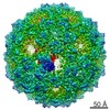


 PDBj
PDBj
 集合体
集合体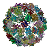
 試料調製
試料調製 電子顕微鏡撮影
 FIELD EMISSION GUN / 加速電圧: 300 kV / 照射モード: FLOOD BEAM
 解析
解析