 Movie
Movie Controller

[English] 日本語
 Yorodumi
- PDB-5a79: Novel inter-subunit contacts in Barley Stripe Mosaic Virus reveal... -
+ Open data

- Basic information
| Entry | Database: PDB / ID: 5a79 | ||||||
|---|---|---|---|---|---|---|---|
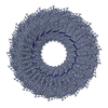
| Title | Novel inter-subunit contacts in Barley Stripe Mosaic Virus revealed by cryo-EM | ||||||
 Components Components |
| ||||||
 Keywords Keywords | VIRUS / BSMV / HELICAL / CRYO-EM / IMAGE PROCESSING / MSA | ||||||
| Function / homology | Tobacco mosaic virus-like, coat protein / Tobacco mosaic virus-like, coat protein superfamily / Virus coat protein (TMV like) / helical viral capsid / structural molecule activity / identical protein binding / RNA / Capsid protein Function and homology information Function and homology information | ||||||
| Biological species |  BARLEY STRIPE MOSAIC VIRUS BARLEY STRIPE MOSAIC VIRUS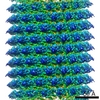 TOBACCO MOSAIC VIRUS TOBACCO MOSAIC VIRUS | ||||||
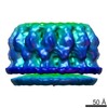
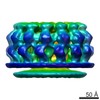
| Method | ELECTRON MICROSCOPY / single particle reconstruction / cryo EM / Resolution: 4.1 Å | ||||||
 Authors Authors | Clare, D.K. / Pechnikova, E. / Skurat, E. / Makarov, V. / Sokolova, O.S. / Solovyev, A.G. / V Orlova, E. | ||||||
 Citation Citation | Journal: Structure / Year: 2015 Title: Novel Inter-Subunit Contacts in Barley Stripe Mosaic Virus Revealed by Cryo-Electron Microscopy. Authors: Daniel Kofi Clare / Eugenia V Pechnikova / Eugene V Skurat / Valentin V Makarov / Olga S Sokolova / Andrey G Solovyev / Elena V Orlova /   Abstract: Barley stripe mosaic virus (BSMV, genus Hordeivirus) is a rod-shaped single-stranded RNA virus similar to viruses of the structurally characterized and well-studied genus Tobamovirus. Here we report ...Barley stripe mosaic virus (BSMV, genus Hordeivirus) is a rod-shaped single-stranded RNA virus similar to viruses of the structurally characterized and well-studied genus Tobamovirus. Here we report the first high-resolution structure of BSMV at 4.1 Å obtained by cryo-electron microscopy. We discovered that BSMV forms two types of virion that differ in the number of coat protein (CP) subunits per turn and interactions between the CP subunits. While BSMV and tobacco mosaic virus CP subunits have a similar fold and interact with RNA using conserved residues, the axial contacts between the CP of these two viral groups are considerably different. BSMV CP subunits lack substantial axial contacts and are held together by a previously unobserved lateral contact formed at the virion surface via an interacting loop, which protrudes from the CP hydrophobic core to the adjacent CP subunit. These data provide an insight into diversity in structural organization of helical viruses. | ||||||
| History |
|
- Structure visualization
| Movie |
Movie viewer |
|---|---|
| Structure viewer | Molecule: MolmilJmol/JSmol |
- Downloads & links
-Download
| PDBx/mmCIF format | 5a79.cif.gz | 50.5 KB | Display | PDBx/mmCIF format |
|---|---|---|---|---|
| PDB format | pdb5a79.ent.gz | 35.6 KB | Display | PDB format |
| PDBx/mmJSON format | 5a79.json.gz | Tree view | PDBx/mmJSON format | |
| Others |  Other downloads Other downloads |
-Validation report
| Arichive directory | https://data.pdbj.org/pub/pdb/validation_reports/a7/5a79ftp://data.pdbj.org/pub/pdb/validation_reports/a7/5a79 | HTTPS FTP |
|---|
-Related structure data
| Related structure data | 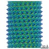 3073MC  3074C 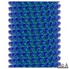 3077C 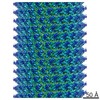 3078C 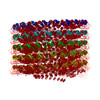 5a7aC M: map data used to model this data C: citing same article ( |
|---|---|
| Similar structure data |


-Links
 PDBj
PDBj
- Assembly
| Deposited unit | 
|
|---|---|
| 1 | x 111
|
| Symmetry | Helical symmetry: (Circular symmetry: 1 / Dyad axis: no / N subunits divisor: 111 / Num. of operations: 111 / Rise per n subunits: 131.2 Å / Rotation per n subunits: 1800 °) |
-Components
| #1: Protein | Mass: 22555.225 Da / Num. of mol.: 1 Source method: isolated from a genetically manipulated source Details: ONLY CONTAINS RESIDUES 3-131 AND 147-194 IN STRUCTURE Source: (gene. exp.) BARLEY STRIPE MOSAIC VIRUSDescription: PURIFIED FROM NICOTIANA BENTHAMIANA INFECTED PLANTS Production host:  AGROBACTERIUM TUMEFACIENS (bacteria) / Strain (production host): GV3101 / References: UniProt: P04866 AGROBACTERIUM TUMEFACIENS (bacteria) / Strain (production host): GV3101 / References: UniProt: P04866 |
|---|---|
| #2: RNA chain | Mass: 958.660 Da / Num. of mol.: 1 / Source method: isolated from a natural source / Source: (natural) TOBACCO MOSAIC VIRUS (STRAIN OM) |
| Sequence details | ONLY CONTAINS RESIDUES 3-131 AND 147-194 IN STRUCTURE |
-Experimental details
-Experiment
| Experiment | Method: ELECTRON MICROSCOPY |
|---|---|
| EM experiment | Aggregation state: FILAMENT / 3D reconstruction method: single particle reconstruction |
- Sample preparation
| Component | Name: CHIMERIC BARLEY STRIPE MOSAIC VIRUS WIDE VIRION / Type: VIRUS |
|---|---|
| Buffer solution | Name: 50MM TRIS-HCL, 50MM KCL, 10MM MGCL2 / pH: 7.5 / Details: 50MM TRIS-HCL, 50MM KCL, 10MM MGCL2 |
| Specimen | Conc.: 3 mg/ml / Embedding applied: NO / Shadowing applied: NO / Staining applied: NO / Vitrification applied: YES |
| Specimen support | Details: CARBON |
| Vitrification | Instrument: HOMEMADE PLUNGER / Cryogen name: ETHANE Details: VITRIFICATION 1 -- CRYOGEN- ETHANE, HUMIDITY- 40, TEMPERATURE- 100, INSTRUMENT- HOMEMADE PLUNGER, METHOD- 3.5UL OF SAMPLE WAS ADDED TO CONTINUOUS CARBON COATED OR C-FLAT GRIDS, BLOTTED FOR 2 ...Details: VITRIFICATION 1 -- CRYOGEN- ETHANE, HUMIDITY- 40, TEMPERATURE- 100, INSTRUMENT- HOMEMADE PLUNGER, METHOD- 3.5UL OF SAMPLE WAS ADDED TO CONTINUOUS CARBON COATED OR C-FLAT GRIDS, BLOTTED FOR 2 SECONDS AND THEN PLUNGED IN LIQUID ETHANE, |
- Electron microscopy imaging
| Experimental equipment |  Model: Tecnai F30 / Image courtesy: FEI Company |
|---|---|
| Microscopy | Model: FEI TECNAI F30 / Date: Jun 1, 2012 |
| Electron gun | Electron source:  FIELD EMISSION GUN / Accelerating voltage: 300 kV / Illumination mode: FLOOD BEAM FIELD EMISSION GUN / Accelerating voltage: 300 kV / Illumination mode: FLOOD BEAM |
| Electron lens | Mode: BRIGHT FIELD / Nominal magnification: 150000 X / Calibrated magnification: 150000 X / Nominal defocus max: 3500 nm / Nominal defocus min: 700 nm / Cs: 2.3 mm |
| Specimen holder | Temperature: 90 K |
| Image recording | Electron dose: 25 e/Å2 / Film or detector model: GATAN ULTRASCAN 4000 (4k x 4k) |
| Image scans | Num. digital images: 297 |
- Processing
| EM software |
| ||||||||||||
|---|---|---|---|---|---|---|---|---|---|---|---|---|---|
| CTF correction | Details: PHASE FLIPPED EACH PARTICLE | ||||||||||||
| 3D reconstruction | Method: PROJECTION MATCHING / Resolution: 4.1 Å / Num. of particles: 2077 / Nominal pixel size: 1 Å / Actual pixel size: 1 Å / Magnification calibration: LAYER LINES / Details: DISORDERED REGIONS WERE MODELED STEREOCHEMICALLY / Symmetry type: HELICAL | ||||||||||||
| Atomic model building | Protocol: FLEXIBLE FIT / Space: REAL / Target criteria: Cross-correlation coefficient Details: METHOD--RIGID BODY USING CHIMERA, FOLLOWED BY FLEXIBLE FITTING IN FLEX-EM AND COOT. THEN REALSPACE REFINEMENT IN PHENIX FOLLOWED BY MANUAL ADJUSTMENT IN COOT (INTERATIVE) REFINEMENT PROTOCOL--CRYO-EM | ||||||||||||
| Refinement | Highest resolution: 4.1 Å | ||||||||||||
| Refinement step | Cycle: LAST / Highest resolution: 4.1 Å
|
