 Movie
Movie Controller


+ Open data

- Basic information
| Entry | Database: PDB / ID: 4v6x | |||||||||
|---|---|---|---|---|---|---|---|---|---|---|
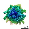
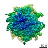
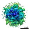
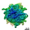
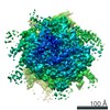
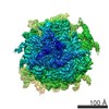
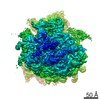
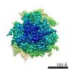
| Title | Structure of the human 80S ribosome | |||||||||
 Components Components |
| |||||||||
 Keywords Keywords | RIBOSOME / eukarya / eukaryotic / ribosomal / 80S / RNA / protein synthesis / mass spectrometry | |||||||||
| Function / homology |  Function and homology information Function and homology informationSynthesis of diphthamide-EEF2 / cytoplasmic translational elongation / translation at postsynapse / ribosome hibernation / translation elongation factor binding / PML body organization / response to folic acid / SUMO binding / translation at presynapse / positive regulation of cytoplasmic translation ...Synthesis of diphthamide-EEF2 / cytoplasmic translational elongation / translation at postsynapse / ribosome hibernation / translation elongation factor binding / PML body organization / response to folic acid / SUMO binding / translation at presynapse / positive regulation of cytoplasmic translation / exit from mitosis / optic nerve development / response to insecticide / regulation of translation involved in cellular response to UV / eukaryotic 80S initiation complex / negative regulation of endoplasmic reticulum unfolded protein response / ribosomal protein import into nucleus / negative regulation of formation of translation preinitiation complex / axial mesoderm development / regulation of G1 to G0 transition / retinal ganglion cell axon guidance / oxidized pyrimidine DNA binding / response to TNF agonist / positive regulation of base-excision repair / positive regulation of ubiquitin-protein transferase activity / positive regulation of respiratory burst involved in inflammatory response / protein-DNA complex disassembly / positive regulation of intrinsic apoptotic signaling pathway in response to DNA damage by p53 class mediator / positive regulation of gastrulation / aggresome / protein tyrosine kinase inhibitor activity / positive regulation of DNA-templated transcription initiation / positive regulation of intrinsic apoptotic signaling pathway in response to DNA damage / 90S preribosome assembly / IRE1-RACK1-PP2A complex / positive regulation of Golgi to plasma membrane protein transport / nucleolus organization / TNFR1-mediated ceramide production / alpha-beta T cell differentiation / positive regulation of DNA damage response, signal transduction by p53 class mediator / GAIT complex / negative regulation of RNA splicing / TORC2 complex binding / neural crest cell differentiation / supercoiled DNA binding / cytoplasmic translational initiation / NF-kappaB complex / negative regulation of DNA repair / G1 to G0 transition / oxidized purine DNA binding / cysteine-type endopeptidase activator activity involved in apoptotic process / middle ear morphogenesis / rRNA modification in the nucleus and cytosol / negative regulation of intrinsic apoptotic signaling pathway in response to hydrogen peroxide / negative regulation of bicellular tight junction assembly / ubiquitin-like protein conjugating enzyme binding / regulation of establishment of cell polarity / negative regulation of phagocytosis / erythrocyte homeostasis / cytoplasmic side of rough endoplasmic reticulum membrane / Formation of the ternary complex, and subsequently, the 43S complex / Uptake and function of diphtheria toxin / lncRNA binding / ion channel inhibitor activity / protein kinase A binding / laminin receptor activity / Ribosomal scanning and start codon recognition / pigmentation / homeostatic process / positive regulation of mitochondrial depolarization / Translation initiation complex formation / macrophage chemotaxis / lung morphogenesis / negative regulation of Wnt signaling pathway / positive regulation of natural killer cell proliferation / male meiosis I / fibroblast growth factor binding / Protein hydroxylation / monocyte chemotaxis / BH3 domain binding / negative regulation of translational frameshifting / regulation of adenylate cyclase-activating G protein-coupled receptor signaling pathway / SARS-CoV-1 modulates host translation machinery / positive regulation of GTPase activity / TOR signaling / mTORC1-mediated signalling / iron-sulfur cluster binding / Peptide chain elongation / translational elongation / regulation of cell division / cellular response to ethanol / skeletal muscle cell differentiation / Selenocysteine synthesis / Formation of a pool of free 40S subunits / negative regulation of protein binding / positive regulation of intrinsic apoptotic signaling pathway by p53 class mediator / Eukaryotic Translation Termination / protein kinase activator activity / protein serine/threonine kinase inhibitor activity / blastocyst development Similarity search - Function | |||||||||
| Biological species |  Homo sapiens (human) Homo sapiens (human) | |||||||||
| Method | ELECTRON MICROSCOPY / single particle reconstruction / cryo EM / Resolution: 5 Å | |||||||||
 Authors Authors | Anger, A.M. / Armache, J.-P. / Berninghausen, O. / Habeck, M. / Subklewe, M. / Wilson, D.N. / Beckmann, R. | |||||||||
 Citation Citation | Journal: Nature / Year: 2013 Title: Structures of the human and Drosophila 80S ribosome. Authors: Andreas M Anger / Jean-Paul Armache / Otto Berninghausen / Michael Habeck / Marion Subklewe / Daniel N Wilson / Roland Beckmann /  Abstract: Protein synthesis in all cells is carried out by macromolecular machines called ribosomes. Although the structures of prokaryotic, yeast and protist ribosomes have been determined, the more complex ...Protein synthesis in all cells is carried out by macromolecular machines called ribosomes. Although the structures of prokaryotic, yeast and protist ribosomes have been determined, the more complex molecular architecture of metazoan 80S ribosomes has so far remained elusive. Here we present structures of Drosophila melanogaster and Homo sapiens 80S ribosomes in complex with the translation factor eEF2, E-site transfer RNA and Stm1-like proteins, based on high-resolution cryo-electron-microscopy density maps. These structures not only illustrate the co-evolution of metazoan-specific ribosomal RNA with ribosomal proteins but also reveal the presence of two additional structural layers in metazoan ribosomes, a well-ordered inner layer covered by a flexible RNA outer layer. The human and Drosophila ribosome structures will provide the basis for more detailed structural, biochemical and genetic experiments. | |||||||||
| History |
|
- Structure visualization
| Movie |
Movie viewer |
|---|---|
| Structure viewer | Molecule: MolmilJmol/JSmol |
- Downloads & links
-Download
| PDBx/mmCIF format | 4v6x.cif.gz | 5.4 MB | Display | PDBx/mmCIF format |
|---|---|---|---|---|
| PDB format | pdb4v6x.ent.gz | Display | PDB format | |
| PDBx/mmJSON format | 4v6x.json.gz | Tree view | PDBx/mmJSON format | |
| Others |  Other downloads Other downloads |
-Validation report
| Arichive directory | https://data.pdbj.org/pub/pdb/validation_reports/v6/4v6xftp://data.pdbj.org/pub/pdb/validation_reports/v6/4v6x | HTTPS FTP |
|---|
-Related structure data
| Related structure data |  5592MC  5591C 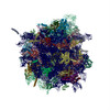 4v6wC M: map data used to model this data C: citing same article ( |
|---|---|
| Similar structure data |


-Links
 PDBj
PDBj


- Assembly
| Deposited unit | 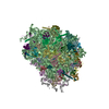
|
|---|---|
| 1 |
|
-Components
-Protein , 3 types, 3 molecules AzAgAh
| #1: Protein | Mass: 95463.211 Da / Num. of mol.: 1 / Source method: isolated from a natural source / Source: (natural) Homo sapiens (human) / References: UniProt: P13639 |
|---|---|
| #2: Protein | Mass: 35115.652 Da / Num. of mol.: 1 / Source method: isolated from a natural source / Source: (natural) Homo sapiens (human) / References: UniProt: P63244 |
| #35: Protein | Mass: 45051.504 Da / Num. of mol.: 1 / Source method: isolated from a natural source / Source: (natural) Homo sapiens (human) / References: UniProt: Q8NC51 |
+40S ribosomal protein ... , 32 types, 32 molecules AUAKAOAXAMASAdANALARAPATABAAAVAYAZAaAbAcADAeAfAJAEACAGAFAHAWAIAQ
-RNA chain , 5 types, 5 molecules B2BCA5A7A8
| #36: RNA chain | Mass: 602776.875 Da / Num. of mol.: 1 / Source method: isolated from a natural source / Source: (natural) Homo sapiens (human) / References: GenBank: X03205 |
|---|---|
| #37: RNA chain | Mass: 24231.510 Da / Num. of mol.: 1 / Source method: isolated from a natural source / Source: (natural) Homo sapiens (human) / References: GenBank: K00328 |
| #85: RNA chain | Mass: 1640222.125 Da / Num. of mol.: 1 / Source method: isolated from a natural source / Source: (natural) Homo sapiens (human) / References: GenBank: NR_003287 |
| #86: RNA chain | Mass: 38998.078 Da / Num. of mol.: 1 / Source method: isolated from a natural source / Source: (natural) Homo sapiens (human) / References: GenBank: V00589 |
| #87: RNA chain | Mass: 50449.812 Da / Num. of mol.: 1 / Source method: isolated from a natural source / Source: (natural) Homo sapiens (human) / References: GenBank: NR_046235 |
+60S ribosomal protein ... , 44 types, 44 molecules CzCKCOCLCVCMCaCNCICDCQCRCACSCTCPCUCXCYCWCZCrChCbCBCFCcCdCeCf...
-60S acidic ribosomal protein ... , 3 types, 5 molecules CqCsCtCuCv
| #39: Protein | Mass: 34309.418 Da / Num. of mol.: 1 / Source method: isolated from a natural source / Source: (natural) Homo sapiens (human) / References: UniProt: P05388 | ||
|---|---|---|---|
| #83: Protein | Mass: 11521.863 Da / Num. of mol.: 2 / Source method: isolated from a natural source / Source: (natural) Homo sapiens (human) / References: UniProt: P05386#84: Protein | Mass: 11676.896 Da / Num. of mol.: 2 / Source method: isolated from a natural source / Source: (natural) Homo sapiens (human) / References: UniProt: P05387 |
-Details
| Has protein modification | Y |
|---|
-Experimental details
-Experiment
| Experiment | Method: ELECTRON MICROSCOPY |
|---|---|
| EM experiment | Aggregation state: PARTICLE / 3D reconstruction method: single particle reconstruction |
- Sample preparation
| Component | Name: 80S human ribosome from PBMCs / Type: RIBOSOME |
|---|---|
| Molecular weight | Value: 4.5 MDa / Experimental value: NO |
| Buffer solution | pH: 7.5 |
| Specimen | Embedding applied: NO / Shadowing applied: NO / Staining applied: NO / Vitrification applied: YES |
| Vitrification | Instrument: FEI VITROBOT MARK IV / Cryogen name: ETHANE / Humidity: 100 % Details: Blot for 3 seconds using two pieces of filter paper, then plunge into liquid ethane (FEI VITROBOT MARK IV). |
- Electron microscopy imaging
| Experimental equipment |  Model: Titan Krios / Image courtesy: FEI Company |
|---|---|
| Microscopy | Model: FEI TITAN KRIOS / Date: Feb 11, 2011 |
| Electron gun | Electron source:  FIELD EMISSION GUN / Accelerating voltage: 200 kV / Illumination mode: FLOOD BEAM FIELD EMISSION GUN / Accelerating voltage: 200 kV / Illumination mode: FLOOD BEAM |
| Electron lens | Mode: BRIGHT FIELD / Nominal magnification: 90000 X / Calibrated magnification: 90000 X / Nominal defocus max: 3500 nm / Nominal defocus min: 800 nm / Cs: 2.7 mm |
| Specimen holder | Specimen holder model: FEI TITAN KRIOS AUTOGRID HOLDER / Specimen holder type: FEI TITAN KRIOS AUTOGRID HOLDER |
| Image recording | Electron dose: 20 e/Å2 / Film or detector model: FEI EAGLE (4k x 4k) |
| Radiation | Protocol: SINGLE WAVELENGTH / Monochromatic (M) / Laue (L): M / Scattering type: x-ray |
| Radiation wavelength | Relative weight: 1 |
- Processing
| EM software | Name: SPIDER / Category: 3D reconstruction | ||||||||||||
|---|---|---|---|---|---|---|---|---|---|---|---|---|---|
| CTF correction | Details: each subvolume | ||||||||||||
| Symmetry | Point symmetry: C1 (asymmetric) | ||||||||||||
| 3D reconstruction | Method: Projection matching / Resolution: 5 Å / Resolution method: FSC 0.5 CUT-OFF / Num. of particles: 343343 / Nominal pixel size: 1.2375 Å / Actual pixel size: 1.2375 Å / Symmetry type: POINT | ||||||||||||
| Refinement step | Cycle: LAST
|
