 Movie
Movie Controller

[English] 日本語
 Yorodumi
- PDB-4v6t: Structure of the bacterial ribosome complexed by tmRNA-SmpB and E... -
+ Open data

- Basic information
| Entry | Database: PDB / ID: 4v6t | ||||||||||||
|---|---|---|---|---|---|---|---|---|---|---|---|---|---|
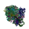
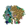
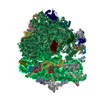
| Title | Structure of the bacterial ribosome complexed by tmRNA-SmpB and EF-G during translocation and MLD-loading | ||||||||||||
 Components Components |
| ||||||||||||
 Keywords Keywords | RIBOSOME / trans-translation / MLD-loading / translocation | ||||||||||||
| Function / homology |  Function and homology information Function and homology informationstringent response / transcription antitermination factor activity, RNA binding / ornithine decarboxylase inhibitor activity / misfolded RNA binding / Group I intron splicing / RNA folding / translational termination / transcriptional attenuation / endoribonuclease inhibitor activity / positive regulation of ribosome biogenesis ...stringent response / transcription antitermination factor activity, RNA binding / ornithine decarboxylase inhibitor activity / misfolded RNA binding / Group I intron splicing / RNA folding / translational termination / transcriptional attenuation / endoribonuclease inhibitor activity / positive regulation of ribosome biogenesis / RNA-binding transcription regulator activity / four-way junction DNA binding / negative regulation of cytoplasmic translation / DnaA-L2 complex / regulation of mRNA stability / translation repressor activity / negative regulation of translational initiation / negative regulation of DNA-templated DNA replication initiation / mRNA regulatory element binding translation repressor activity / regulation of DNA-templated transcription elongation / positive regulation of RNA splicing / transcription elongation factor complex / response to reactive oxygen species / cytosolic ribosome assembly / ribosome assembly / assembly of large subunit precursor of preribosome / transcription antitermination / DNA endonuclease activity / regulation of cell growth / DNA-templated transcription termination / response to radiation / maintenance of translational fidelity / mRNA 5'-UTR binding / regulation of translation / large ribosomal subunit / transferase activity / ribosomal small subunit assembly / ribosome biogenesis / ribosome binding / ribosomal small subunit biogenesis / 5S rRNA binding / ribosomal large subunit assembly / small ribosomal subunit / small ribosomal subunit rRNA binding / cytosolic small ribosomal subunit / large ribosomal subunit rRNA binding / cytosolic large ribosomal subunit / cytoplasmic translation / tRNA binding / negative regulation of translation / rRNA binding / structural constituent of ribosome / ribosome / translation / response to antibiotic / negative regulation of DNA-templated transcription / hydrolase activity / mRNA binding / DNA binding / RNA binding / zinc ion binding / membrane / cytoplasm / cytosol Similarity search - Function | ||||||||||||
| Biological species |  | ||||||||||||
| Method | ELECTRON MICROSCOPY / single particle reconstruction / cryo EM / Resolution: 8.3 Å | ||||||||||||
 Authors Authors | Ramrath, D.J.F. / Yamamoto, H. / Rother, K. / Wittek, D. / Pech, M. / Mielke, T. / Loerke, J. / Scheerer, P. / Ivanov, P. / Teraoka, Y. ...Ramrath, D.J.F. / Yamamoto, H. / Rother, K. / Wittek, D. / Pech, M. / Mielke, T. / Loerke, J. / Scheerer, P. / Ivanov, P. / Teraoka, Y. / Shpanchenko, O. / Nierhaus, K.H. / Spahn, C.M.T. | ||||||||||||
 Citation Citation | Journal: Nature / Year: 2012 Title: The complex of tmRNA-SmpB and EF-G on translocating ribosomes. Authors: David J F Ramrath / Hiroshi Yamamoto / Kristian Rother / Daniela Wittek / Markus Pech / Thorsten Mielke / Justus Loerke / Patrick Scheerer / Pavel Ivanov / Yoshika Teraoka / Olga Shpanchenko ...Authors: David J F Ramrath / Hiroshi Yamamoto / Kristian Rother / Daniela Wittek / Markus Pech / Thorsten Mielke / Justus Loerke / Patrick Scheerer / Pavel Ivanov / Yoshika Teraoka / Olga Shpanchenko / Knud H Nierhaus / Christian M T Spahn /  Abstract: Bacterial ribosomes stalled at the 3' end of malfunctioning messenger RNAs can be rescued by transfer-messenger RNA (tmRNA)-mediated trans-translation. The SmpB protein forms a complex with the ...Bacterial ribosomes stalled at the 3' end of malfunctioning messenger RNAs can be rescued by transfer-messenger RNA (tmRNA)-mediated trans-translation. The SmpB protein forms a complex with the tmRNA, and the transfer-RNA-like domain (TLD) of the tmRNA then enters the A site of the ribosome. Subsequently, the TLD-SmpB module is translocated to the P site, a process that is facilitated by the elongation factor EF-G, and translation is switched to the mRNA-like domain (MLD) of the tmRNA. Accurate loading of the MLD into the mRNA path is an unusual initiation mechanism. Despite various snapshots of different ribosome-tmRNA complexes at low to intermediate resolution, it is unclear how the large, highly structured tmRNA is translocated and how the MLD is loaded. Here we present a cryo-electron microscopy reconstruction of a fusidic-acid-stalled ribosomal 70S-tmRNA-SmpB-EF-G complex (carrying both of the large ligands, that is, EF-G and tmRNA) at 8.3 Å resolution. This post-translocational intermediate (TI(POST)) presents the TLD-SmpB module in an intrasubunit ap/P hybrid site and a tRNA(fMet) in an intrasubunit pe/E hybrid site. Conformational changes in the ribosome and tmRNA occur in the intersubunit space and on the solvent side. The key underlying event is a unique extra-large swivel movement of the 30S head, which is crucial for both tmRNA-SmpB translocation and MLD loading, thereby coupling translocation to MLD loading. This mechanism exemplifies the versatile, dynamic nature of the ribosome, and it shows that the conformational modes of the ribosome that normally drive canonical translation can also be used in a modified form to facilitate more complex tasks in specialized non-canonical pathways. | ||||||||||||
| History |
|
- Structure visualization
| Movie |
Movie viewer |
|---|---|
| Structure viewer | Molecule: MolmilJmol/JSmol |
- Downloads & links
-Download
| PDBx/mmCIF format | 4v6t.cif.gz | 3.5 MB | Display | PDBx/mmCIF format |
|---|---|---|---|---|
| PDB format | pdb4v6t.ent.gz | Display | PDB format | |
| PDBx/mmJSON format | 4v6t.json.gz | Tree view | PDBx/mmJSON format | |
| Others |  Other downloads Other downloads |
-Validation report
| Arichive directory | https://data.pdbj.org/pub/pdb/validation_reports/v6/4v6tftp://data.pdbj.org/pub/pdb/validation_reports/v6/4v6t | HTTPS FTP |
|---|
-Related structure data
| Related structure data |  5386MC M: map data used to model this data C: citing same article ( |
|---|---|
| Similar structure data |


-Links
 PDBj
PDBj


- Assembly
| Deposited unit | 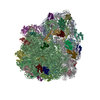
|
|---|---|
| 1 | 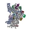
|
-Components
-RNA chain , 5 types, 5 molecules AAAVAXBABB
| #1: RNA chain | Mass: 498725.406 Da / Num. of mol.: 1 / Source method: isolated from a natural source / Source: (natural) |
|---|---|
| #22: RNA chain | Mass: 117212.297 Da / Num. of mol.: 1 / Source method: isolated from a natural source / Source: (natural) |
| #24: RNA chain | Mass: 24802.785 Da / Num. of mol.: 1 / Source method: isolated from a natural source / Source: (natural) |
| #26: RNA chain | Mass: 941306.188 Da / Num. of mol.: 1 / Source method: isolated from a natural source / Source: (natural) |
| #56: RNA chain | Mass: 38483.926 Da / Num. of mol.: 1 / Source method: isolated from a natural source / Source: (natural) |
-30S ribosomal protein ... , 20 types, 20 molecules ABACADAEAFAGAHAIAJAKALAMANAOAPAQARASATAU
| #2: Protein | Mass: 24253.943 Da / Num. of mol.: 1 / Source method: isolated from a natural source / Source: (natural) |
|---|---|
| #3: Protein | Mass: 23078.785 Da / Num. of mol.: 1 / Source method: isolated from a natural source / Source: (natural) |
| #4: Protein | Mass: 23383.002 Da / Num. of mol.: 1 / Source method: isolated from a natural source / Source: (natural) |
| #5: Protein | Mass: 15804.282 Da / Num. of mol.: 1 / Source method: isolated from a natural source / Source: (natural) |
| #6: Protein | Mass: 11669.371 Da / Num. of mol.: 1 / Source method: isolated from a natural source / Source: (natural) |
| #7: Protein | Mass: 16861.523 Da / Num. of mol.: 1 / Source method: isolated from a natural source / Source: (natural) |
| #8: Protein | Mass: 14015.361 Da / Num. of mol.: 1 / Source method: isolated from a natural source / Source: (natural) |
| #9: Protein | Mass: 14554.882 Da / Num. of mol.: 1 / Source method: isolated from a natural source / Source: (natural) |
| #10: Protein | Mass: 11196.988 Da / Num. of mol.: 1 / Source method: isolated from a natural source / Source: (natural) |
| #11: Protein | Mass: 12487.200 Da / Num. of mol.: 1 / Source method: isolated from a natural source / Source: (natural) |
| #12: Protein | Mass: 13636.961 Da / Num. of mol.: 1 / Source method: isolated from a natural source / Source: (natural) |
| #13: Protein | Mass: 12625.753 Da / Num. of mol.: 1 / Source method: isolated from a natural source / Source: (natural) |
| #14: Protein | Mass: 11475.364 Da / Num. of mol.: 1 / Source method: isolated from a natural source / Source: (natural) |
| #15: Protein | Mass: 10159.621 Da / Num. of mol.: 1 / Source method: isolated from a natural source / Source: (natural) |
| #16: Protein | Mass: 9207.572 Da / Num. of mol.: 1 / Source method: isolated from a natural source / Source: (natural) |
| #17: Protein | Mass: 9263.946 Da / Num. of mol.: 1 / Source method: isolated from a natural source / Source: (natural) |
| #18: Protein | Mass: 6466.477 Da / Num. of mol.: 1 / Source method: isolated from a natural source / Source: (natural) |
| #19: Protein | Mass: 9057.626 Da / Num. of mol.: 1 / Source method: isolated from a natural source / Source: (natural) |
| #20: Protein | Mass: 9506.190 Da / Num. of mol.: 1 / Source method: isolated from a natural source / Source: (natural) |
| #21: Protein | Mass: 6067.081 Da / Num. of mol.: 1 / Source method: isolated from a natural source / Source: (natural) |
-Protein , 2 types, 2 molecules AWAY
| #23: Protein | Mass: 14177.479 Da / Num. of mol.: 1 / Fragment: SEE REMARK 999 Source method: isolated from a genetically manipulated source Source: (gene. exp.) |
|---|---|
| #25: Protein | Mass: 76977.102 Da / Num. of mol.: 1 / Fragment: SEE REMARK 999 Source method: isolated from a genetically manipulated source Source: (gene. exp.) |
+50S ribosomal protein ... , 29 types, 29 molecules BCBDBEBFBGBHBIBJBKBLBMBNBOBPBQBRBSBTBUBVBWBXBYBZB0B1B2B3B4
-Details
| Sequence details | MODELED SEQUENCES FOR SSRA_BINDING PROTEIN (UNP Q8RR57 RESIDUES 1-123) AND ELONGATION FACTOR G (UNP ...MODELED SEQUENCES FOR SSRA_BINDING PROTEIN (UNP Q8RR57 RESIDUES 1-123) AND ELONGATION |
|---|
-Experimental details
-Experiment
| Experiment | Method: ELECTRON MICROSCOPY |
|---|---|
| EM experiment | Aggregation state: PARTICLE / 3D reconstruction method: single particle reconstruction |
- Sample preparation
| Component | Name: Ribosomal complex 70S-tmRNA-SmpB-tRNA-EFG-GDP-FA / Type: RIBOSOME / Details: in vitro reconstituted ribosomal complex |
|---|---|
| Buffer solution | pH: 7.6 |
| Specimen | Conc.: 0.15 mg/ml / Embedding applied: NO / Shadowing applied: NO / Staining applied: NO / Vitrification applied: YES |
| Specimen support | Details: Quantifoil R3-3 Cu mesh with 2 nm carbon support film |
| Vitrification | Instrument: FEI VITROBOT MARK I / Cryogen name: ETHANE / Temp: 96.15 K / Humidity: 100 % Details: blot for 5-10 seconds before plunging into liquid ethane (FEI Vitrobot) Method: blot for 5-10 seconds before plunging |
- Electron microscopy imaging
| Experimental equipment |  Model: Tecnai Polara / Image courtesy: FEI Company |
|---|---|
| Microscopy | Model: FEI POLARA 300 / Date: Dec 8, 2009 |
| Electron gun | Electron source:  FIELD EMISSION GUN / Accelerating voltage: 300 kV / Illumination mode: SPOT SCAN FIELD EMISSION GUN / Accelerating voltage: 300 kV / Illumination mode: SPOT SCAN |
| Electron lens | Mode: BRIGHT FIELD / Nominal magnification: 39000 X / Calibrated magnification: 39000 X / Nominal defocus max: 5000 nm / Nominal defocus min: 2500 nm / Cs: 2 mm |
| Specimen holder | Temperature: 77 K |
| Image recording | Electron dose: 20 e/Å2 / Film or detector model: KODAK SO-163 FILM |
| Image scans | Num. digital images: 302 |
| Radiation | Protocol: SINGLE WAVELENGTH / Monochromatic (M) / Laue (L): M / Scattering type: x-ray |
| Radiation wavelength | Relative weight: 1 |
- Processing
| EM software |
| ||||||||||||
|---|---|---|---|---|---|---|---|---|---|---|---|---|---|
| CTF correction | Details: The volumes were CTF-corrected in defocus groups, with an average of approximately 215 individual images per group | ||||||||||||
| Symmetry | Point symmetry: C1 (asymmetric) | ||||||||||||
| 3D reconstruction | Method: angular refinement / Resolution: 8.3 Å / Num. of particles: 68843 / Nominal pixel size: 1.26 Å / Actual pixel size: 1.26 Å / Symmetry type: POINT | ||||||||||||
| Refinement step | Cycle: LAST
|
