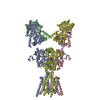
Movie
Movie Controller


+ Open data

- Basic information
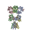
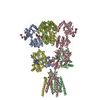
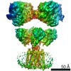
| Entry | Database: PDB / ID: 4uqj | ||||||
|---|---|---|---|---|---|---|---|
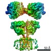
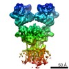
| Title | Cryo-EM density map of GluA2em in complex with ZK200775 | ||||||
 Components Components | GLUTAMATE RECEPTOR 2 | ||||||
 Keywords Keywords | TRANSPORT PROTEIN / GLUA2EM ANTAGONIST-BOUND CLOSED STATE | ||||||
| Function / homology |  Function and homology information Function and homology informationspine synapse / dendritic spine neck / dendritic spine cytoplasm / dendritic spine head / cellular response to amine stimulus / Activation of AMPA receptors / ligand-gated monoatomic cation channel activity / perisynaptic space / Trafficking of GluR2-containing AMPA receptors / response to lithium ion ...spine synapse / dendritic spine neck / dendritic spine cytoplasm / dendritic spine head / cellular response to amine stimulus / Activation of AMPA receptors / ligand-gated monoatomic cation channel activity / perisynaptic space / Trafficking of GluR2-containing AMPA receptors / response to lithium ion / AMPA glutamate receptor activity / AMPA glutamate receptor clustering / kainate selective glutamate receptor activity / immunoglobulin binding / AMPA glutamate receptor complex / regulation of receptor recycling / extracellularly glutamate-gated ion channel activity / cellular response to glycine / ionotropic glutamate receptor complex / asymmetric synapse / Unblocking of NMDA receptors, glutamate binding and activation / glutamate receptor binding / conditioned place preference / positive regulation of synaptic transmission / response to fungicide / regulation of synaptic transmission, glutamatergic / extracellular ligand-gated monoatomic ion channel activity / cytoskeletal protein binding / glutamate-gated receptor activity / cellular response to brain-derived neurotrophic factor stimulus / regulation of long-term synaptic depression / somatodendritic compartment / glutamate-gated calcium ion channel activity / presynaptic active zone membrane / excitatory synapse / ionotropic glutamate receptor signaling pathway / ionotropic glutamate receptor binding / dendrite membrane / dendrite cytoplasm / ligand-gated monoatomic ion channel activity involved in regulation of presynaptic membrane potential / positive regulation of excitatory postsynaptic potential / dendritic shaft / SNARE binding / synaptic membrane / PDZ domain binding / establishment of protein localization / protein tetramerization / transmitter-gated monoatomic ion channel activity involved in regulation of postsynaptic membrane potential / synaptic transmission, glutamatergic / cerebral cortex development / receptor internalization / postsynaptic density membrane / modulation of chemical synaptic transmission / Schaffer collateral - CA1 synapse / terminal bouton / synaptic vesicle / long-term synaptic potentiation / amyloid-beta binding / synaptic vesicle membrane / growth cone / presynapse / signaling receptor activity / scaffold protein binding / presynaptic membrane / dendritic spine / chemical synaptic transmission / perikaryon / postsynaptic membrane / neuron projection / postsynaptic density / external side of plasma membrane / axon / neuronal cell body / synapse / dendrite / protein kinase binding / protein-containing complex binding / glutamatergic synapse / cell surface / endoplasmic reticulum / protein-containing complex / membrane / identical protein binding / plasma membrane Similarity search - Function | ||||||
| Biological species |  | ||||||
| Method | ELECTRON MICROSCOPY / single particle reconstruction / cryo EM / Resolution: 10.4 Å | ||||||
 Authors Authors | Meyerson, J.R. / Kumar, J. / Chittori, S. / Rao, P. / Pierson, J. / Bartesaghi, A. / Mayer, M.L. / Subramaniam, S. | ||||||
 Citation Citation | Journal: Nature / Year: 2014 Title: Structural mechanism of glutamate receptor activation and desensitization. Authors: Joel R Meyerson / Janesh Kumar / Sagar Chittori / Prashant Rao / Jason Pierson / Alberto Bartesaghi / Mark L Mayer / Sriram Subramaniam /  Abstract: Ionotropic glutamate receptors are ligand-gated ion channels that mediate excitatory synaptic transmission in the vertebrate brain. To gain a better understanding of how structural changes gate ion ...Ionotropic glutamate receptors are ligand-gated ion channels that mediate excitatory synaptic transmission in the vertebrate brain. To gain a better understanding of how structural changes gate ion flux across the membrane, we trapped rat AMPA (α-amino-3-hydroxy-5-methyl-4-isoxazole propionic acid) and kainate receptor subtypes in their major functional states and analysed the resulting structures using cryo-electron microscopy. We show that transition to the active state involves a 'corkscrew' motion of the receptor assembly, driven by closure of the ligand-binding domain. Desensitization is accompanied by disruption of the amino-terminal domain tetramer in AMPA, but not kainate, receptors with a two-fold to four-fold symmetry transition in the ligand-binding domains in both subtypes. The 7.6 Å structure of a desensitized kainate receptor shows how these changes accommodate channel closing. These findings integrate previous physiological, biochemical and structural analyses of glutamate receptors and provide a molecular explanation for key steps in receptor gating. | ||||||
| History |
|
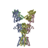
- Structure visualization
| Movie |
Movie viewer |
|---|---|
| Structure viewer | Molecule: MolmilJmol/JSmol |
- Downloads & links
-Download
| PDBx/mmCIF format | 4uqj.cif.gz | 480.8 KB | Display | PDBx/mmCIF format |
|---|---|---|---|---|
| PDB format | pdb4uqj.ent.gz | 374.2 KB | Display | PDB format |
| PDBx/mmJSON format | 4uqj.json.gz | Tree view | PDBx/mmJSON format | |
| Others |  Other downloads Other downloads |
-Validation report
| Arichive directory | https://data.pdbj.org/pub/pdb/validation_reports/uq/4uqjftp://data.pdbj.org/pub/pdb/validation_reports/uq/4uqj | HTTPS FTP |
|---|
-Related structure data
| Related structure data | 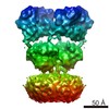 2680MC 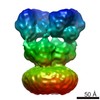 2684C 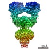 2685C 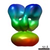 2686C 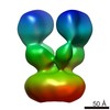 2687C 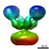 2688C 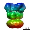 2689C 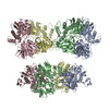 4uq6C 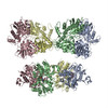 4uqkC 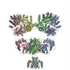 4uqqC C: citing same article ( M: map data used to model this data |
|---|---|
| Similar structure data |
-Links
 PDBj
PDBj


- Assembly
| Deposited unit | 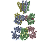
|
|---|---|
| 1 |
|
-Components
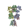
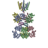
| #1: Protein | Mass: 92523.617 Da / Num. of mol.: 4 / Fragment: RESIDUES 22-847 / Mutation: YES Source method: isolated from a genetically manipulated source Source: (gene. exp.)   SPODOPTERA FRUGIPERDA (fall armyworm) / References: UniProt: P19491 SPODOPTERA FRUGIPERDA (fall armyworm) / References: UniProt: P19491#2: Chemical | ChemComp-ZK1 / {[ 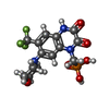  Mass: 409.254 Da / Num. of mol.: 4 / Source method: obtained synthetically / Formula: C14H15F3N3O6P / Comment: medication*YM Mass: 409.254 Da / Num. of mol.: 4 / Source method: obtained synthetically / Formula: C14H15F3N3O6P / Comment: medication*YMHas protein modification | Y | |
|---|
-Experimental details
-Experiment
| Experiment | Method: ELECTRON MICROSCOPY |
|---|---|
| EM experiment | Aggregation state: PARTICLE / 3D reconstruction method: single particle reconstruction |
- Sample preparation
| Component | Name: GLUA2 / Type: COMPLEX |
|---|---|
| Buffer solution | Name: 150 MM NACL, 20 MM TRIS, 0.75 MM DDM, 0.12 MM CHS, 0.3 MM ZK200775 pH: 8 Details: 150 MM NACL, 20 MM TRIS, 0.75 MM DDM, 0.12 MM CHS, 0.3 MM ZK200775 |
| Specimen | Conc.: 1.8 mg/ml / Embedding applied: NO / Shadowing applied: NO / Staining applied: NO / Vitrification applied: YES |
| Specimen support | Details: HOLEY CARBON |
| Vitrification | Instrument: FEI VITROBOT MARK IV / Cryogen name: ETHANE / Details: ETHANE |
- Electron microscopy imaging
| Experimental equipment |  Model: Titan Krios / Image courtesy: FEI Company |
|---|---|
| Microscopy | Model: FEI TITAN KRIOS / Date: Aug 1, 2013 |
| Electron gun | Electron source:  FIELD EMISSION GUN / Accelerating voltage: 300 kV / Illumination mode: FLOOD BEAM FIELD EMISSION GUN / Accelerating voltage: 300 kV / Illumination mode: FLOOD BEAM |
| Electron lens | Mode: BRIGHT FIELD / Nominal magnification: 47000 X / Nominal defocus max: 3500 nm / Nominal defocus min: 2000 nm |
| Image recording | Electron dose: 25 e/Å2 / Film or detector model: FEI FALCON II (4k x 4k) |
- Processing
| EM software |
| ||||||||||||
|---|---|---|---|---|---|---|---|---|---|---|---|---|---|
| CTF correction | Details: INDIVIDUAL PARTICLES | ||||||||||||
| Symmetry | Point symmetry: C2 (2 fold cyclic) | ||||||||||||
| 3D reconstruction | Resolution: 10.4 Å / Num. of particles: 26795 Details: COORDINATES FROM 3KG2 WERE FIT AS FIVE INDEPENDENT RIGID BODIES CONSISTING OF TWO ATD DIMERS AND TWO LBD DIMERS AND THE TMD. GEOMETRY AND STEREOCHEMISTRY OUTLIERS ARE THOSE PRESENT IN PDB ...Details: COORDINATES FROM 3KG2 WERE FIT AS FIVE INDEPENDENT RIGID BODIES CONSISTING OF TWO ATD DIMERS AND TWO LBD DIMERS AND THE TMD. GEOMETRY AND STEREOCHEMISTRY OUTLIERS ARE THOSE PRESENT IN PDB 3KG2 USED FOR RIGID BODY FITS SUBMISSION BASED ON EXPERIMENTAL DATA FROM EMDB EMD-2680. (DEPOSITION ID: 12574). Symmetry type: POINT | ||||||||||||
| Atomic model building | Protocol: RIGID BODY FIT / Space: REAL / Details: METHOD--RIGID BODY REFINEMENT PROTOCOL--X-RAY | ||||||||||||
| Atomic model building | PDB-ID: 3KG2 Accession code: 3KG2 / Source name: PDB / Type: experimental model | ||||||||||||
| Refinement | Highest resolution: 10.4 Å | ||||||||||||
| Refinement step | Cycle: LAST / Highest resolution: 10.4 Å
|