 ムービー
ムービー コントローラー


+ データを開く

- 基本情報
基本情報
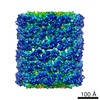
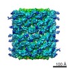
| 登録情報 | データベース: PDB / ID: 4bed | |||||||||
|---|---|---|---|---|---|---|---|---|---|---|
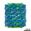
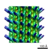
| タイトル | Keyhole limpet hemocyanin (KLH): 9A cryoEM structure and molecular model of the KLH1 didecamer reveal the interfaces and intricate topology of the 160 functional units | |||||||||
 要素 要素 | (HEMOCYANIN KLH1) x 2 | |||||||||
 キーワード キーワード | OXYGEN TRANSPORT / KEYHOLE LIMPET HEMOCYANIN / KLH / GASTROPODA / OXYGEN CARRIER | |||||||||
| 機能・相同性 |  機能・相同性情報 機能・相同性情報oxygen carrier activity / oxidoreductase activity / extracellular region / metal ion binding 類似検索 - 分子機能 | |||||||||
| 生物種 |  MEGATHURA CRENULATA (無脊椎動物) MEGATHURA CRENULATA (無脊椎動物) | |||||||||
| 手法 | 電子顕微鏡法 / 単粒子再構成法 / クライオ電子顕微鏡法 / 解像度: 9 Å | |||||||||
 データ登録者 データ登録者 | Gatsogiannis, C. / Markl, J. | |||||||||
 引用 引用 | ジャーナル: J Mol Biol / 年: 2009 タイトル: Keyhole limpet hemocyanin: 9-A CryoEM structure and molecular model of the KLH1 didecamer reveal the interfaces and intricate topology of the 160 functional units. 著者: Christos Gatsogiannis / Jürgen Markl /  要旨: Hemocyanins are blue copper-containing respiratory proteins in the hemolymph of many arthropods and molluscs. Molluscan hemocyanins are decamers, didecamers, or multidecamers of a 340- to 400-kDa ...Hemocyanins are blue copper-containing respiratory proteins in the hemolymph of many arthropods and molluscs. Molluscan hemocyanins are decamers, didecamers, or multidecamers of a 340- to 400-kDa polypeptide subunit containing seven or eight globular functional units (FUs; FU-a to FU-h), each with an oxygen-binding site. The decamers are short 35-nm hollow cylinders, with their lumen narrowed by a collar complex. Our recently published 9-A cryo-electron microscopy/crystal structure hybrid model of a 3.4-MDa cephalopod hemocyanin decamer [Nautilus pompilius hemocyanin (NpH)] revealed the pathway of the seven-FU subunit (340 kDa), 15 types of inter-FU interface, and an asymmetric collar consisting of five "arcs" (FU-g pairs). We now present a comparable hybrid model of an 8-MDa gastropod hemocyanin didecamer assembled from two asymmetric decamers [isoform keyhole limpet hemocyanin (KLH) 1 of the established immunogen KLH]. Compared to NpH, the KLH1 subunit (400 kDa) is C-terminally elongated by FU-h, which is further extended by a unique tail domain. We have found that the wall-and-arc structure of the KLH1 decamer is very similar to that of NpH. We have traced the subunit pathway and how it continues from KLH1-g to KLH1-h to form an annulus of five "slabs" (FU-h pairs) at one cylinder edge. The 15 types of inter-FU interface detected in NpH are also present in KLH1. Moreover, we have identified one arc/slab interface, two slab/slab interfaces, five slab/wall interfaces, and four decamer/decamer interfaces. The 27 interfaces are described on the basis of two subunit conformers, yielding an asymmetric homodimer. Six protrusions from the cryo-electron microscopy structure per subunit are associated with putative attachment sites for N-linked glycans, indicating a total of 120 sugar trees in KLH1. Also, putative binding sites for divalent cations have been detected. In conclusion, the present 9-A data on KLH1 confirm and substantially broaden our recent analysis of the smaller cephalopod hemocyanin and essentially solve the gastropod hemocyanin structure. #1: ジャーナル: Biochim Biophys Acta / 年: 2013 タイトル: Evolution of molluscan hemocyanin structures. 著者: Jürgen Markl / 要旨: Hemocyanin transports oxygen in the hemolymph of many molluscs and arthropods and is therefore a central physiological factor in these animals. Molluscan hemocyanin molecules are oligomers composed ...Hemocyanin transports oxygen in the hemolymph of many molluscs and arthropods and is therefore a central physiological factor in these animals. Molluscan hemocyanin molecules are oligomers composed of many protein subunits that in turn encompass subsets of distinct functional units. The structure and evolution of molluscan hemocyanin have been studied for decades, but it required the recent progress in DNA sequencing, X-ray crystallography and 3D electron microscopy to produce a detailed view of their structure and evolution. The basic quaternary structure is a cylindrical decamer 35nm in diameter, consisting of wall and collar (typically at one end of the cylinder). Depending on the animal species, decamers, didecamers and multidecamers occur in the hemolymph. Whereas the wall architecture of the decamer seems to be invariant, four different types of collar have been identified in different molluscan taxa. Correspondingly, there exist four subunit types that differ in their collar functional units and range from 350 to 550kDa. Thus, molluscan hemocyanin subunits are among the largest polypeptides in nature. In this report, recent 3D reconstructions are used to explain and visualize the different functional units, subunits and quaternary structures of molluscan hemocyanins. Moreover, on the basis of DNA analyses and structural considerations, their possible evolution is traced. This article is part of a Special Issue entitled: Oxygen Binding and Sensing Proteins. | |||||||||
| 履歴 |
|
- 構造の表示
構造の表示
| ムービー |
ムービービューア |
|---|---|
| 構造ビューア | 分子: MolmilJmol/JSmol |
- ダウンロードとリンク
-ダウンロード
| PDBx/mmCIF形式 | 4bed.cif.gz | 1.2 MB | 表示 | PDBx/mmCIF形式 |
|---|---|---|---|---|
| PDB形式 | pdb4bed.ent.gz | 954.5 KB | 表示 | PDB形式 |
| PDBx/mmJSON形式 | 4bed.json.gz | ツリー表示 | PDBx/mmJSON形式 | |
| その他 |  その他のダウンロード その他のダウンロード |
-検証レポート
| アーカイブディレクトリ | https://data.pdbj.org/pub/pdb/validation_reports/be/4bedftp://data.pdbj.org/pub/pdb/validation_reports/be/4bed | HTTPS FTP |
|---|
-関連構造データ
-リンク
 PDBj
PDBj- 集合体
集合体
| 登録構造単位 | 
|
|---|---|
| 1 | x 10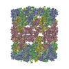
|
| 2 |
|
| 3 | 
|
| 対称性 | 点対称性: (シェーンフリース記号: D (2回xn回 2面回転対称)) |
-要素
| #1: タンパク質 | 分子量: 191623.828 Da / 分子数: 2 / 由来タイプ: 天然 / 由来: (天然) MEGATHURA CRENULATA (無脊椎動物) / 参照: UniProt: Q53IP9, UniProt: Q10583*PLUS#2: タンパク質 | 分子量: 199504.094 Da / 分子数: 2 / 由来タイプ: 天然 / 由来: (天然) MEGATHURA CRENULATA (無脊椎動物) / 参照: UniProt: Q53IP9, UniProt: Q10583*PLUS#3: 化合物 | ChemComp-CUO /   分子量: 159.091 Da / 分子数: 16 / 由来タイプ: 合成 / 式: Cu2O2 分子量: 159.091 Da / 分子数: 16 / 由来タイプ: 合成 / 式: Cu2O2Has protein modification | Y | 配列の詳細 | THIS PDB ENTRY IS BASED ON AN UPDATED SEQUENCE | |
|---|
-実験情報
-実験
| 実験 | 手法: 電子顕微鏡法 |
|---|---|
| EM実験 | 試料の集合状態: PARTICLE / 3次元再構成法: 単粒子再構成法 |
- 試料調製
試料調製
| 構成要素 | 名称: KEYHOLE LIMPET HEMOCYANIN ISOFORM 1 (KLH1) / タイプ: COMPLEX |
|---|---|
| 緩衝液 | 名称: 50 MM TRIS-HCL, 150 MM NACL, 5MM CACL, 5MM MGCL2 / pH: 7.4 / 詳細: 50 MM TRIS-HCL, 150 MM NACL, 5MM CACL, 5MM MGCL2 |
| 試料 | 濃度: 0.7 mg/ml / 包埋: NO / シャドウイング: NO / 染色: NO / 凍結: YES |
| 試料支持 | 詳細: HOLEY CARBON |
| 急速凍結 | 装置: HOMEMADE PLUNGER / 凍結剤: ETHANE 詳細: VITRIFICATION INSTRUMENT - HOME MADE. VITRIFICATION CARRIED OUT IN 25 PERCENT OXYGEN ATMOSPHERE. SINGLE- SIDED BLOTTING |
- 電子顕微鏡撮影
| 実験機器 |  モデル: Tecnai F20 / 画像提供: FEI Company |
|---|---|
| 顕微鏡 | モデル: FEI TECNAI F20 / 日付: 2007年7月25日 |
| 電子銃 | 電子線源:  FIELD EMISSION GUN / 加速電圧: 200 kV / 照射モード: FLOOD BEAM FIELD EMISSION GUN / 加速電圧: 200 kV / 照射モード: FLOOD BEAM |
| 電子レンズ | モード: BRIGHT FIELD / 倍率(公称値): 50000 X / 倍率(補正後): 50000 X / 最大 デフォーカス(公称値): 4000 nm / 最小 デフォーカス(公称値): 800 nm / Cs: 2 mm |
| 試料ホルダ | 温度: 68 K |
| 撮影 | フィルム・検出器のモデル: KODAK SO-163 FILM |
| 画像スキャン | デジタル画像の数: 98 |
- 解析
解析
| EMソフトウェア | 名称: IMAGIC / バージョン: 5 / カテゴリ: 3次元再構成 | ||||||||||||
|---|---|---|---|---|---|---|---|---|---|---|---|---|---|
| CTF補正 | 詳細: PER MICROGRAPH | ||||||||||||
| 対称性 | 点対称性: D5 (2回x5回 2面回転対称) | ||||||||||||
| 3次元再構成 | 解像度: 9 Å / 粒子像の数: 4762 / ピクセルサイズ(公称値): 1 Å / ピクセルサイズ(実測値): 1 Å 詳細: KLH1 IS A CYLINDRICAL 8 MDA PROTEIN WITH D5 SYMMETRY AND COMPOSED OF 20 IDENTICAL 400 KDA POLYPEPTIDE SUBUNITS. THE ASYMMETRIC UNIT IS A SUBUNIT DIMER (800 KDA). THE TWO CONSTITUENT ...詳細: KLH1 IS A CYLINDRICAL 8 MDA PROTEIN WITH D5 SYMMETRY AND COMPOSED OF 20 IDENTICAL 400 KDA POLYPEPTIDE SUBUNITS. THE ASYMMETRIC UNIT IS A SUBUNIT DIMER (800 KDA). THE TWO CONSTITUENT POLYPEPTIDES ARE REPRESENTED HERE BY FOUR CHAINS (A, B, C, D). THIS ALLOWS A QUICK VISUALIZATION OF TWO ALTERNATIVE SUBUNIT PATHWAYS THAT HAVE BEEN PROPOSED: A-B + C-D (THIS STUDY) AND A-D + C-B (PDB-ID 3J32). THE 400 KDA POLYPEPTIDE IS FOLDED INTO EIGHT FUNCTIONAL UNITS, TERMED KLH1-A TO KLH1-H (1-421, 422-835, 836-1255, 1256-1664, 1665-2081, 2082-2488, 2489-2905, 2906-3398). THEY ARE CONCATENATED VIA LONG LINKERS. FOR THIS MODEL, THE STRUCTURE OF KLH1-H WAS AVAILABLE (PDB-ID 3QJO). HOMOLOGY MODELING OF THE OTHER FUNCTIONAL UNITS WAS DONE USING PDB-ID 1JS8 AS TEMPLATE. EACH FUNCTIONAL UNIT IS ABLE TO BIND A DIOXYGEN MOLECULE BETWEEN TWO COPPER IONS COORDINATED BY SIX HISTIDINES: 42, 60, 69, 179, 183, 210; 462, 482, 491, 602, 606, 633; 876, 896, 905, 1015, 1019, 1046; 1293, 1311, 1320, 1424, 1428, 1455; 1705, 1725, 1734, 1847, 1851, 1878; 2122, 2141, 2150, 2260, 2264, 2291; 2542, 2561, 2570, 2670, 2674, 2701; 2946, 2965, 2974, 3075, 3079, 3106. SUBMISSION BASED ON EXPERIMENTAL DATA FROM EMDB EMD-1569. (DEPOSITION ID: 6428). 対称性のタイプ: POINT | ||||||||||||
| 原子モデル構築 | プロトコル: RIGID BODY FIT / 詳細: METHOD--RIGID-BODY FITTING AND LOOP REFINEMENT | ||||||||||||
| 精密化 | 最高解像度: 9 Å | ||||||||||||
| 精密化ステップ | サイクル: LAST / 最高解像度: 9 Å
|
