[English] 日本語
 Yorodumi
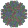
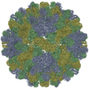
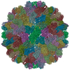
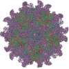
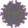
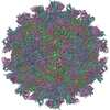
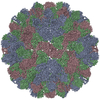
- PDB-3zx8: Cryo-EM reconstruction of native and expanded Turnip Crinkle virus -
+ Open data
- Basic information
| Entry | Database: PDB / ID: 3zx8 | ||||||
|---|---|---|---|---|---|---|---|
| Title | Cryo-EM reconstruction of native and expanded Turnip Crinkle virus | ||||||
 Components Components | CAPSID PROTEIN | ||||||
 Keywords Keywords | VIRUS / GENOMIC RNA STRUCTURE / GENOME UNCOATING / SSRNA VIRUS / ICOSAHEDRAL | ||||||
| Function / homology |  Function and homology information Function and homology informationT=3 icosahedral viral capsid / symbiont-mediated suppression of host innate immune response / : / structural molecule activity / RNA binding Similarity search - Function | ||||||
| Biological species |  TURNIP CRINKLE VIRUS TURNIP CRINKLE VIRUS | ||||||
| Method | ELECTRON MICROSCOPY / single particle reconstruction / cryo EM / Resolution: 11.5 Å | ||||||
 Authors Authors | Bakker, S.E. / Robottom, J. / Hogle, J.M. / Maeda, A. / Pearson, A.R. / Stockley, P.G. / Ranson, N.A. / Harrison, S.C. | ||||||
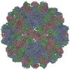
 Citation Citation |  Journal: J Mol Biol / Year: 2012 Journal: J Mol Biol / Year: 2012Title: Isolation of an asymmetric RNA uncoating intermediate for a single-stranded RNA plant virus. Authors: Saskia E Bakker / Robert J Ford / Amy M Barker / Janice Robottom / Keith Saunders / Arwen R Pearson / Neil A Ranson / Peter G Stockley /  Abstract: We have determined the three-dimensional structures of both native and expanded forms of turnip crinkle virus (TCV), using cryo-electron microscopy, which allows direct visualization of the ...We have determined the three-dimensional structures of both native and expanded forms of turnip crinkle virus (TCV), using cryo-electron microscopy, which allows direct visualization of the encapsidated single-stranded RNA and coat protein (CP) N-terminal regions not seen in the high-resolution X-ray structure of the virion. The expanded form, which is a putative disassembly intermediate during infection, arises from a separation of the capsid-forming domains of the CP subunits. Capsid expansion leads to the formation of pores that could allow exit of the viral RNA. A subset of the CP N-terminal regions becomes proteolytically accessible in the expanded form, although the RNA remains inaccessible to nuclease. Sedimentation velocity assays suggest that the expanded state is metastable and that expansion is not fully reversible. Proteolytically cleaved CP subunits dissociate from the capsid, presumably leading to increased electrostatic repulsion within the viral RNA. Consistent with this idea, electron microscopy images show that proteolysis introduces asymmetry into the TCV capsid and allows initial extrusion of the genome from a defined site. The apparent formation of polysomes in wheat germ extracts suggests that subsequent uncoating is linked to translation. The implication is that the viral RNA and its capsid play multiple roles during primary infections, consistent with ribosome-mediated genome uncoating to avoid host antiviral activity. #1:  Journal: J Mol Biol / Year: 1986 Journal: J Mol Biol / Year: 1986Title: Structure and assembly of turnip crinkle virus. I. X-ray crystallographic structure analysis at 3.2 A resolution. Abstract: The structure of turnip crinkle virus has been determined at 3.2 A resolution, using the electron density of tomato bushy stunt virus as a starting point for phase refinement by non-crystallographic ...The structure of turnip crinkle virus has been determined at 3.2 A resolution, using the electron density of tomato bushy stunt virus as a starting point for phase refinement by non-crystallographic symmetry. The structures are very closely related, especially in the subunit arm and S domain, where only small insertions and deletions and small co-ordinate shifts relate one chain to another. The P domains, although quite similar in fold, are oriented somewhat differently with respect to the S domains. Understanding of the structure of turnip crinkle virus has been important for analyzing its assembly, as described in an accompanying paper. | ||||||
| History |
| ||||||
| Remark 650 | HELIX DETERMINATION METHOD: AUTHOR PROVIDED. | ||||||
| Remark 700 | SHEET DETERMINATION METHOD: AUTHOR PROVIDED. |
- Structure visualization
| Movie |
 Movie viewer Movie viewer |
|---|---|
| Structure viewer | Molecule:  Molmil Molmil Jmol/JSmol Jmol/JSmol |
- Downloads & links
- Download
| PDBx/mmCIF format |  3zx8.cif.gz 3zx8.cif.gz | 161.2 KB | Display |  PDBx/mmCIF format PDBx/mmCIF format |
|---|---|---|---|---|
| PDB format |  pdb3zx8.ent.gz pdb3zx8.ent.gz | 114.6 KB | Display |  PDB format PDB format |
| PDBx/mmJSON format |  3zx8.json.gz 3zx8.json.gz | Tree view |  PDBx/mmJSON format PDBx/mmJSON format | |
| Others |  Other downloads Other downloads |
-Validation report
| Summary document |  3zx8_validation.pdf.gz 3zx8_validation.pdf.gz | 807.3 KB | Display |  wwPDB validaton report wwPDB validaton report |
|---|---|---|---|---|
| Full document |  3zx8_full_validation.pdf.gz 3zx8_full_validation.pdf.gz | 1.1 MB | Display | |
| Data in XML |  3zx8_validation.xml.gz 3zx8_validation.xml.gz | 81.7 KB | Display | |
| Data in CIF |  3zx8_validation.cif.gz 3zx8_validation.cif.gz | 102.2 KB | Display | |
| Arichive directory |  https://data.pdbj.org/pub/pdb/validation_reports/zx/3zx8 https://data.pdbj.org/pub/pdb/validation_reports/zx/3zx8 ftp://data.pdbj.org/pub/pdb/validation_reports/zx/3zx8 ftp://data.pdbj.org/pub/pdb/validation_reports/zx/3zx8 | HTTPS FTP |
-Related structure data
| Related structure data |  1863MC  1864C  3zx9C C: citing same article ( M: map data used to model this data |
|---|---|
| Similar structure data |
- Links
Links
- Assembly
| Deposited unit | 
|
|---|---|
| 1 | x 60
|
| 2 |
|
| 3 | x 5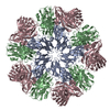
|
| 4 | x 6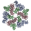
|
| 5 | 
|
| Symmetry | Point symmetry: (Schoenflies symbol: I (icosahedral)) |
- Components
| #1: Protein | Mass: 37755.586 Da / Num. of mol.: 3 / Source method: isolated from a natural source / Source: (natural)  TURNIP CRINKLE VIRUS / Strain: M / References: UniProt: P06663 TURNIP CRINKLE VIRUS / Strain: M / References: UniProt: P06663Sequence details | IN CHAIN C RESIDUES 1-52 DISORDERED AND NOT OBSERVED. IN CHAINS A AND B RESIDUES 1-80 DISORDERED ...IN CHAIN C RESIDUES 1-52 DISORDERED | |
|---|
-Experimental details
-Experiment
| Experiment | Method: ELECTRON MICROSCOPY |
|---|---|
| EM experiment | Aggregation state: PARTICLE / 3D reconstruction method: single particle reconstruction |
- Sample preparation
| Component | Name: TURNIP CRINKLE VIRUS / Type: VIRUS |
|---|---|
| Buffer solution | Name: 10 MM SODIUM PHOSPHATE PH 7.4, 10 MM MAGNESIUM SULPHATE pH: 7.4 Details: 10 MM SODIUM PHOSPHATE PH 7.4, 10 MM MAGNESIUM SULPHATE |
| Specimen | Conc.: 2 mg/ml / Embedding applied: NO / Shadowing applied: NO / Staining applied: NO / Vitrification applied: YES |
| Specimen support | Details: HOLEY CARBON |
| Vitrification | Instrument: HOMEMADE PLUNGER / Cryogen name: ETHANE Details: VITRIFICATION 1 -- CRYOGEN- ETHANE, TEMPERATURE- 77, INSTRUMENT- DOUBLE SIDED AUTOMATED BLOTTER AND PLUNGER, METHOD- BLOT 1.6 SECONDS BEFORE PLUNGING, |
- Electron microscopy imaging
| Experimental equipment |  Model: Tecnai F20 / Image courtesy: FEI Company |
|---|---|
| Microscopy | Model: FEI TECNAI F20 |
| Electron gun | Electron source:  FIELD EMISSION GUN / Accelerating voltage: 200 kV / Illumination mode: FLOOD BEAM FIELD EMISSION GUN / Accelerating voltage: 200 kV / Illumination mode: FLOOD BEAM |
| Electron lens | Mode: BRIGHT FIELD / Nominal magnification: 50000 X / Calibrated magnification: 52911 X / Nominal defocus max: 3500 nm / Nominal defocus min: 1000 nm / Cs: 2 mm |
| Image recording | Electron dose: 15 e/Å2 / Film or detector model: KODAK SO-163 FILM |
| Image scans | Num. digital images: 70 |
- Processing
| EM software | Name: SPIDER / Category: 3D reconstruction | ||||||||||||
|---|---|---|---|---|---|---|---|---|---|---|---|---|---|
| CTF correction | Details: PHASE-FLIPPING EACH PARTICLE | ||||||||||||
| Symmetry | Point symmetry: I (icosahedral) | ||||||||||||
| 3D reconstruction | Method: ICOSAHEDRAL / Resolution: 11.5 Å / Num. of particles: 18681 / Nominal pixel size: 1.4 Å / Actual pixel size: 1.323 Å Details: SUBMISSION BASED ON EXPERIMENTAL DATA FROM EMDB EMD-1863. (DEPOSITION ID: 7817). Symmetry type: POINT | ||||||||||||
| Atomic model building | Protocol: RIGID BODY FIT / Space: REAL / Details: METHOD--RIGID-BODY FIT REFINEMENT PROTOCOL--X-RAY | ||||||||||||
| Refinement | Highest resolution: 11.5 Å | ||||||||||||
| Refinement step | Cycle: LAST / Highest resolution: 11.5 Å
|
 Movie
Movie Controller




 PDBj
PDBj

