Movie
Movie Controller

[English] 日本語
 Yorodumi
- PDB-3jc2: The structure of the mammalian Sec61 channel opened by a signal s... -
+ Open data

- Basic information
| Entry | Database: PDB / ID: 3jc2 | ||||||
|---|---|---|---|---|---|---|---|
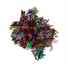
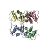
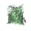
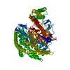
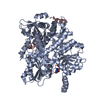
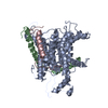
| Title | The structure of the mammalian Sec61 channel opened by a signal sequence | ||||||
 Components Components |
| ||||||
 Keywords Keywords | TRANSPORT PROTEIN / Sec61 / translocation / signal sequence | ||||||
| Function / homology |  Function and homology information Function and homology informationlong-day photoperiodism / response to external biotic stimulus / prolactin receptor binding / positive regulation of biosynthetic process / peptide hormone secretion / Insertion of tail-anchored proteins into the endoplasmic reticulum membrane / membrane docking / pronephric nephron development / response to L-arginine / biosynthetic process ...long-day photoperiodism / response to external biotic stimulus / prolactin receptor binding / positive regulation of biosynthetic process / peptide hormone secretion / Insertion of tail-anchored proteins into the endoplasmic reticulum membrane / membrane docking / pronephric nephron development / response to L-arginine / biosynthetic process / regulation of meiotic cell cycle process involved in oocyte maturation / cotranslational protein targeting to membrane / Ssh1 translocon complex / Sec61 translocon complex / protein insertion into ER membrane / positive regulation of lactation / protein targeting to ER / SRP-dependent cotranslational protein targeting to membrane, translocation / positive regulation of fatty acid biosynthetic process / signal sequence receptor activity / mammary gland development / post-translational protein targeting to membrane, translocation / blastocyst formation / response to food / transmembrane protein transporter activity / positive regulation of endocytosis / response to mechanical stimulus / lactation / positive regulation of phagocytosis / positive regulation of receptor signaling pathway via JAK-STAT / female pregnancy / hormone activity / response to nutrient levels / positive regulation of non-canonical NF-kappaB signal transduction / phospholipid binding / positive regulation of nitric oxide biosynthetic process / ribosome binding / positive regulation of canonical NF-kappaB signal transduction / cell surface receptor signaling pathway / endoplasmic reticulum lumen / inflammatory response / negative regulation of gene expression / positive regulation of gene expression / endoplasmic reticulum membrane / negative regulation of apoptotic process / : / extracellular region / metal ion binding / cytosol Similarity search - Function | ||||||
| Biological species |  | ||||||
| Method | ELECTRON MICROSCOPY / single particle reconstruction / cryo EM / Resolution: 3.6 Å | ||||||
 Authors Authors | Voorhees, R.M. / Hegde, R.S. | ||||||
 Citation Citation | Journal: Science / Year: 2016 Title: Structure of the Sec61 channel opened by a signal sequence. Authors: Rebecca M Voorhees / Ramanujan S Hegde /  Abstract: Secreted and integral membrane proteins compose up to one-third of the biological proteome. These proteins contain hydrophobic signals that direct their translocation across or insertion into the ...Secreted and integral membrane proteins compose up to one-third of the biological proteome. These proteins contain hydrophobic signals that direct their translocation across or insertion into the lipid bilayer by the Sec61 protein-conducting channel. The molecular basis of how hydrophobic signals within a nascent polypeptide trigger channel opening is not understood. Here, we used cryo-electron microscopy to determine the structure of an active Sec61 channel that has been opened by a signal sequence. The signal supplants helix 2 of Sec61α, which triggers a rotation that opens the central pore both axially across the membrane and laterally toward the lipid bilayer. Comparisons with structures of Sec61 in other states suggest a pathway for how hydrophobic signals engage the channel to gain access to the lipid bilayer. | ||||||
| History |
|
- Structure visualization
| Movie |
Movie viewer |
|---|---|
| Structure viewer | Molecule: MolmilJmol/JSmol |
- Downloads & links
-Download
| PDBx/mmCIF format | 3jc2.cif.gz | 110.1 KB | Display | PDBx/mmCIF format |
|---|---|---|---|---|
| PDB format | pdb3jc2.ent.gz | 85 KB | Display | PDB format |
| PDBx/mmJSON format | 3jc2.json.gz | Tree view | PDBx/mmJSON format | |
| Others |  Other downloads Other downloads |
-Validation report
| Arichive directory | https://data.pdbj.org/pub/pdb/validation_reports/jc/3jc2ftp://data.pdbj.org/pub/pdb/validation_reports/jc/3jc2 | HTTPS FTP |
|---|
-Related structure data
| Related structure data | 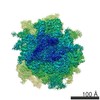 3245MC M: map data used to model this data C: citing same article ( |
|---|---|
| Similar structure data |

-Links
 PDBj
PDBj


- Assembly
| Deposited unit | 
|
|---|---|
| 1 |
|
-Components
| #1: Protein | Mass: 52279.379 Da / Num. of mol.: 1 / Source method: isolated from a natural source / Details: purified from native canine microsomes / Source: (natural) |
|---|---|
| #2: Protein | Mass: 7019.456 Da / Num. of mol.: 1 / Source method: isolated from a natural source / Details: purified from native canine microsomes / Source: (natural) |
| #3: Protein/peptide | Mass: 2053.576 Da / Num. of mol.: 1 Source method: isolated from a genetically manipulated source Source: (gene. exp.) Tissue fraction (production host): reticulocyte translation extract in the presence of canine microsomes References: UniProt: Q6VMP1, UniProt: P01239*PLUS |
| #4: Protein/peptide | Mass: 2741.370 Da / Num. of mol.: 1 / Source method: isolated from a natural source / Details: purified from native canine microsomes / Source: (natural) |
| Has protein modification | N |
-Experimental details
-Experiment
| Experiment | Method: ELECTRON MICROSCOPY |
|---|---|
| EM experiment | Aggregation state: PARTICLE / 3D reconstruction method: single particle reconstruction |
- Sample preparation
| Component |
| ||||||||||||||||||||||||
|---|---|---|---|---|---|---|---|---|---|---|---|---|---|---|---|---|---|---|---|---|---|---|---|---|---|
| Buffer solution | Name: 50 mM HEPES, 200 mM potassium acetate, 15 mM magnesium acetate, 1 mM DTT, 0.25% Digitonin pH: 7.5 Details: 50 mM HEPES, 200 mM potassium acetate, 15 mM magnesium acetate, 1 mM DTT, 0.25% Digitonin | ||||||||||||||||||||||||
| Specimen | Embedding applied: NO / Shadowing applied: NO / Staining applied: NO / Vitrification applied: YES | ||||||||||||||||||||||||
| Specimen support | Details: Quantifoil (R2/2) holey carbon grid covered in a 70 Angstrom-thick layer of amorphous carbon | ||||||||||||||||||||||||
| Vitrification | Instrument: FEI VITROBOT MARK I / Cryogen name: ETHANE / Humidity: 100 % Details: 3 uL sample was added to the grid, incubated for 30 seconds at 4 C, blotted for 9 seconds, and then plunged into liquid ethane (FEI VITROBOT). Method: 3 uL sample was added to the grid, incubated for 30 seconds at 4 C, blotted for 9 seconds |
- Electron microscopy imaging
| Experimental equipment |  Model: Titan Krios / Image courtesy: FEI Company |
|---|---|
| Microscopy | Model: FEI TITAN KRIOS / Date: Mar 6, 2015 |
| Electron gun | Electron source:  FIELD EMISSION GUN / Accelerating voltage: 300 kV / Illumination mode: SPOT SCAN FIELD EMISSION GUN / Accelerating voltage: 300 kV / Illumination mode: SPOT SCAN |
| Electron lens | Mode: BRIGHT FIELD / Nominal magnification: 59000 X / Calibrated magnification: 104478 X / Nominal defocus max: 3500 nm / Nominal defocus min: 2000 nm |
| Image recording | Electron dose: 27 e/Å2 / Film or detector model: FEI FALCON II (4k x 4k) |
- Processing
| EM software |
| |||||||||||||||||||||
|---|---|---|---|---|---|---|---|---|---|---|---|---|---|---|---|---|---|---|---|---|---|---|
| Symmetry | Point symmetry: C1 (asymmetric) | |||||||||||||||||||||
| 3D reconstruction | Resolution: 3.6 Å / Num. of particles: 101339 / Nominal pixel size: 1.34 Å / Actual pixel size: 1.34 Å / Symmetry type: POINT | |||||||||||||||||||||
| Atomic model building | Space: REAL / Target criteria: R-factor Details: DETAILS--Real-space fitting using Chimera and Coot followed by reciprocal-space refinement using REFMAC | |||||||||||||||||||||
| Atomic model building |
| |||||||||||||||||||||
| Refinement step | Cycle: LAST
|