 Movie
Movie Controller


+ Open data

- Basic information
| Entry | Database: PDB / ID: 3j9x | ||||||
|---|---|---|---|---|---|---|---|
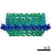
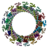
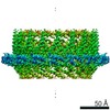
| Title | A Virus that Infects a Hyperthermophile Encapsidates A-Form DNA | ||||||
 Components Components |
| ||||||
 Keywords Keywords | VIRUS / archaeal virus / helical symmetry | ||||||
| Function / homology |  Function and homology information Function and homology information | ||||||
| Biological species |   Sulfolobus islandicus rod-shaped virus 2 Sulfolobus islandicus rod-shaped virus 2 | ||||||
| Method | ELECTRON MICROSCOPY / helical reconstruction / cryo EM / Resolution: 3.8 Å | ||||||
 Authors Authors | DiMaio, F. / Yu, X. / Rensen, E. / Krupovic, M. / Prangishvili, D. / Egelman, E. | ||||||
 Citation Citation | Journal: Science / Year: 2015 Title: Virology. A virus that infects a hyperthermophile encapsidates A-form DNA. Authors: Frank DiMaio / Xiong Yu / Elena Rensen / Mart Krupovic / David Prangishvili / Edward H Egelman /   Abstract: Extremophiles, microorganisms thriving in extreme environmental conditions, must have proteins and nucleic acids that are stable at extremes of temperature and pH. The nonenveloped, rod-shaped virus ...Extremophiles, microorganisms thriving in extreme environmental conditions, must have proteins and nucleic acids that are stable at extremes of temperature and pH. The nonenveloped, rod-shaped virus SIRV2 (Sulfolobus islandicus rod-shaped virus 2) infects the hyperthermophilic acidophile Sulfolobus islandicus, which lives at 80°C and pH 3. We have used cryo-electron microscopy to generate a three-dimensional reconstruction of the SIRV2 virion at ~4 angstrom resolution, which revealed a previously unknown form of virion organization. Although almost half of the capsid protein is unstructured in solution, this unstructured region folds in the virion into a single extended α helix that wraps around the DNA. The DNA is entirely in the A-form, which suggests a common mechanism with bacterial spores for protecting DNA in the most adverse environments. | ||||||
| History |
|
- Structure visualization
| Movie |
Movie viewer |
|---|---|
| Structure viewer | Molecule: MolmilJmol/JSmol |
- Downloads & links
-Download
| PDBx/mmCIF format | 3j9x.cif.gz | 1.4 MB | Display | PDBx/mmCIF format |
|---|---|---|---|---|
| PDB format | pdb3j9x.ent.gz | 1.1 MB | Display | PDB format |
| PDBx/mmJSON format | 3j9x.json.gz | Tree view | PDBx/mmJSON format | |
| Others |  Other downloads Other downloads |
-Validation report
| Summary document | 3j9x_validation.pdf.gz | 1.2 MB | Display | wwPDB validaton report |
|---|---|---|---|---|
| Full document | 3j9x_full_validation.pdf.gz | 1.2 MB | Display | |
| Data in XML | 3j9x_validation.xml.gz | 154.1 KB | Display | |
| Data in CIF | 3j9x_validation.cif.gz | 239 KB | Display | |
| Arichive directory | https://data.pdbj.org/pub/pdb/validation_reports/j9/3j9xftp://data.pdbj.org/pub/pdb/validation_reports/j9/3j9x | HTTPS FTP |
-Related structure data
| Related structure data | 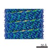 6310MC M: map data used to model this data C: citing same article ( |
|---|---|
| Similar structure data | |
| EM raw data | EMPIAR-10060 (Title: Subset of image stack used for 3D reconstruction / Data size: 35.9 Data #1: image segments of SIRV2 [micrographs - single frame]) |


-Links
 PDBj
PDBj


- Assembly
| Deposited unit | 
|
|---|---|
| 1 |
|
| Symmetry | Helical symmetry: (Circular symmetry: 1 / Dyad axis: no / N subunits divisor: 1 / Num. of operations: 30 / Rise per n subunits: 2.9 Å / Rotation per n subunits: 24.5 °) |
| Details | The helical parameters generate the capsid filament from chains A and B. |
-Components
| #1: Protein | Mass: 13760.465 Da / Num. of mol.: 58 / Source method: isolated from a natural source / Source: (natural) Sulfolobus islandicus rod-shaped virus 2 / References: UniProt: Q8V9P2#2: DNA chain | Mass: 107382.531 Da / Num. of mol.: 2 / Source method: isolated from a natural source / Source: (natural) Sulfolobus islandicus rod-shaped virus 2 |
|---|
-Experimental details
-Experiment
| Experiment | Method: ELECTRON MICROSCOPY |
|---|---|
| EM experiment | Aggregation state: FILAMENT / 3D reconstruction method: helical reconstruction |
- Sample preparation
| Component | Name: Sulfolobus islandicus rod-shaped virus 2 / Type: VIRUS / Synonym: SIRV2 |
|---|---|
| Details of virus | Empty: NO / Enveloped: NO / Host category: ARCHAEA / Isolate: SPECIES / Type: VIRION |
| Natural host | Organism: Sulfolobus islandicus |
| Buffer solution | pH: 6 |
| Specimen | Embedding applied: NO / Shadowing applied: NO / Staining applied: NO / Vitrification applied: YES |
| Vitrification | Instrument: FEI VITROBOT MARK IV / Cryogen name: ETHANE / Humidity: 100 % / Details: Plunged into liquid ethane (FEI VITROBOT MARK IV). |
- Electron microscopy imaging
| Experimental equipment |  Model: Titan Krios / Image courtesy: FEI Company |
|---|---|
| Microscopy | Model: FEI TITAN KRIOS / Date: Sep 19, 2014 |
| Electron gun | Electron source:  FIELD EMISSION GUN / Accelerating voltage: 300 kV / Illumination mode: FLOOD BEAM FIELD EMISSION GUN / Accelerating voltage: 300 kV / Illumination mode: FLOOD BEAM |
| Electron lens | Mode: BRIGHT FIELD / Nominal defocus max: 3000 nm / Nominal defocus min: 500 nm / Cs: 2.7 mm |
| Specimen holder | Specimen holder model: FEI TITAN KRIOS AUTOGRID HOLDER |
| Image recording | Electron dose: 20 e/Å2 / Film or detector model: FEI FALCON II (4k x 4k) |
| Image scans | Num. digital images: 437 |
| Radiation | Protocol: SINGLE WAVELENGTH / Monochromatic (M) / Laue (L): M / Scattering type: x-ray |
| Radiation wavelength | Relative weight: 1 |
- Processing
| EM software | Name: SPIDER / Category: 3D reconstruction | ||||||||||||
|---|---|---|---|---|---|---|---|---|---|---|---|---|---|
| CTF correction | Details: multiply images by CTF, divide reconstruction by sum of CTF**2 | ||||||||||||
| Helical symmerty | Angular rotation/subunit: 24.5 ° / Axial rise/subunit: 2.9 Å / Axial symmetry: C1 | ||||||||||||
| 3D reconstruction | Method: IHRSR / Resolution: 3.8 Å / Resolution method: FSC 0.143 CUT-OFF / Nominal pixel size: 1.05 Å / Actual pixel size: 1.05 Å / Details: (Helical Details: IHRSR) / Symmetry type: HELICAL | ||||||||||||
| Refinement step | Cycle: LAST
|
