[English] 日本語
 Yorodumi
- PDB-3j4j: Model of full-length T. thermophilus Translation Initiation Facto... -
+ Open data
- Basic information
| Entry | Database: PDB / ID: 3j4j | ||||||
|---|---|---|---|---|---|---|---|
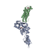
| Title | Model of full-length T. thermophilus Translation Initiation Factor 2 refined against its cryo-EM density from a 30S Initiation Complex map | ||||||
 Components Components | Translation initiation factor IF-2 | ||||||
 Keywords Keywords | TRANSLATION / IF2 / GTP-Binding Protein / fMet-tRNA binding / ribosome binding | ||||||
| Function / homology |  Function and homology information Function and homology informationtranslation initiation factor activity / GTPase activity / GTP binding / cytosol Similarity search - Function | ||||||
| Biological species |   Thermus thermophilus (bacteria) Thermus thermophilus (bacteria) | ||||||
| Method | ELECTRON MICROSCOPY / single particle reconstruction / cryo EM / Resolution: 11.5 Å | ||||||
 Authors Authors | Simonetti, A. / Marzi, S. / Billas, I.M.L. / Tsai, A. / Fabbretti, A. / Myasnikov, A. / Roblin, P. / Vaiana, A.C. / Hazemann, I. / Eiler, D. ...Simonetti, A. / Marzi, S. / Billas, I.M.L. / Tsai, A. / Fabbretti, A. / Myasnikov, A. / Roblin, P. / Vaiana, A.C. / Hazemann, I. / Eiler, D. / Steitz, T.A. / Puglisi, J.D. / Gualerzi, C.O. / Klaholz, B.P. | ||||||
 Citation Citation |  Journal: Proc Natl Acad Sci U S A / Year: 2013 Journal: Proc Natl Acad Sci U S A / Year: 2013Title: Involvement of protein IF2 N domain in ribosomal subunit joining revealed from architecture and function of the full-length initiation factor. Authors: Angelita Simonetti / Stefano Marzi / Isabelle M L Billas / Albert Tsai / Attilio Fabbretti / Alexander G Myasnikov / Pierre Roblin / Andrea C Vaiana / Isabelle Hazemann / Daniel Eiler / ...Authors: Angelita Simonetti / Stefano Marzi / Isabelle M L Billas / Albert Tsai / Attilio Fabbretti / Alexander G Myasnikov / Pierre Roblin / Andrea C Vaiana / Isabelle Hazemann / Daniel Eiler / Thomas A Steitz / Joseph D Puglisi / Claudio O Gualerzi / Bruno P Klaholz /  Abstract: Translation initiation factor 2 (IF2) promotes 30S initiation complex (IC) formation and 50S subunit joining, which produces the 70S IC. The architecture of full-length IF2, determined by small angle ...Translation initiation factor 2 (IF2) promotes 30S initiation complex (IC) formation and 50S subunit joining, which produces the 70S IC. The architecture of full-length IF2, determined by small angle X-ray diffraction and cryo electron microscopy, reveals a more extended conformation of IF2 in solution and on the ribosome than in the crystal. The N-terminal domain is only partially visible in the 30S IC, but in the 70S IC, it stabilizes interactions between IF2 and the L7/L12 stalk of the 50S, and on its deletion, proper N-formyl-methionyl(fMet)-tRNA(fMet) positioning and efficient transpeptidation are affected. Accordingly, fast kinetics and single-molecule fluorescence data indicate that the N terminus promotes 70S IC formation by stabilizing the productive sampling of the 50S subunit during 30S IC joining. Together, our data highlight the dynamics of IF2-dependent ribosomal subunit joining and the role played by the N terminus of IF2 in this process. | ||||||
| History |
|
- Structure visualization
| Movie |
 Movie viewer Movie viewer |
|---|---|
| Structure viewer | Molecule:  Molmil Molmil Jmol/JSmol Jmol/JSmol |
- Downloads & links
- Download
| PDBx/mmCIF format |  3j4j.cif.gz 3j4j.cif.gz | 112.9 KB | Display |  PDBx/mmCIF format PDBx/mmCIF format |
|---|---|---|---|---|
| PDB format |  pdb3j4j.ent.gz pdb3j4j.ent.gz | 80.5 KB | Display |  PDB format PDB format |
| PDBx/mmJSON format |  3j4j.json.gz 3j4j.json.gz | Tree view |  PDBx/mmJSON format PDBx/mmJSON format | |
| Others |  Other downloads Other downloads |
-Validation report
| Summary document |  3j4j_validation.pdf.gz 3j4j_validation.pdf.gz | 653 KB | Display |  wwPDB validaton report wwPDB validaton report |
|---|---|---|---|---|
| Full document |  3j4j_full_validation.pdf.gz 3j4j_full_validation.pdf.gz | 748.3 KB | Display | |
| Data in XML |  3j4j_validation.xml.gz 3j4j_validation.xml.gz | 33.9 KB | Display | |
| Data in CIF |  3j4j_validation.cif.gz 3j4j_validation.cif.gz | 47.1 KB | Display | |
| Arichive directory |  https://data.pdbj.org/pub/pdb/validation_reports/j4/3j4j https://data.pdbj.org/pub/pdb/validation_reports/j4/3j4j ftp://data.pdbj.org/pub/pdb/validation_reports/j4/3j4j ftp://data.pdbj.org/pub/pdb/validation_reports/j4/3j4j | HTTPS FTP |
-Related structure data
| Related structure data |  2448MC M: map data used to model this data C: citing same article ( |
|---|---|
| Similar structure data |
- Links
Links
- Assembly
| Deposited unit | 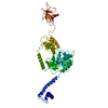
|
|---|---|
| 1 |
|
- Components
| #1: Protein | Mass: 62494.469 Da / Num. of mol.: 1 Source method: isolated from a genetically manipulated source Source: (gene. exp.)   Thermus thermophilus (bacteria) / Strain: HB8 / ATCC 27634 / DSM 579 / Gene: infB, TTHA0699 / Production host: Thermus thermophilus (bacteria) / Strain: HB8 / ATCC 27634 / DSM 579 / Gene: infB, TTHA0699 / Production host:  |
|---|
-Experimental details
-Experiment
| Experiment | Method: ELECTRON MICROSCOPY |
|---|---|
| EM experiment | Aggregation state: PARTICLE / 3D reconstruction method: single particle reconstruction |
- Sample preparation
| Component |
| ||||||||||||
|---|---|---|---|---|---|---|---|---|---|---|---|---|---|
| Buffer solution | Name: 10 mM HEPES, pH 7.5, 70 mM NH4Cl, 30 mM KCl, 8 mM magnesium acetate, 1 mM DTT pH: 7.5 Details: 10 mM HEPES, pH 7.5, 70 mM NH4Cl, 30 mM KCl, 8 mM magnesium acetate, 1 mM DTT | ||||||||||||
| Specimen | Conc.: 0.5 mg/ml / Embedding applied: NO / Shadowing applied: NO / Staining applied: NO / Vitrification applied: YES | ||||||||||||
| Vitrification | Instrument: FEI VITROBOT MARK I / Cryogen name: ETHANE / Details: Vitrification carried out in liquid ethane |
- Electron microscopy imaging
| Experimental equipment |  Model: Tecnai F30 / Image courtesy: FEI Company |
|---|---|
| Microscopy | Model: FEI TECNAI F30 / Date: Oct 9, 2011 |
| Electron gun | Electron source:  FIELD EMISSION GUN / Accelerating voltage: 150 kV / Illumination mode: FLOOD BEAM FIELD EMISSION GUN / Accelerating voltage: 150 kV / Illumination mode: FLOOD BEAM |
| Electron lens | Mode: BRIGHT FIELD / Nominal magnification: 59000 X / Nominal defocus max: -3500 nm / Nominal defocus min: -1500 nm |
| Specimen holder | Temperature: 70 K |
| Image recording | Electron dose: 1.5 e/Å2 / Film or detector model: FEI EAGLE (4k x 4k) |
| Radiation | Protocol: SINGLE WAVELENGTH / Monochromatic (M) / Laue (L): M / Scattering type: x-ray |
| Radiation wavelength | Relative weight: 1 |
- Processing
| EM software |
| ||||||||||||||||
|---|---|---|---|---|---|---|---|---|---|---|---|---|---|---|---|---|---|
| Symmetry | Point symmetry: C1 (asymmetric) | ||||||||||||||||
| 3D reconstruction | Resolution: 11.5 Å / Num. of particles: 13000 / Nominal pixel size: 1.92 Å / Symmetry type: POINT | ||||||||||||||||
| Atomic model building | Protocol: FLEXIBLE FIT / Space: REAL / Target criteria: cross-correlation coefficient Details: METHOD--cross-correlation gradient based REFINEMENT PROTOCOL--flexible fitting, dynamic elastic network | ||||||||||||||||
| Refinement step | Cycle: LAST
|
 Movie
Movie Controller




 PDBj
PDBj


