 Movie
Movie Controller

[English] 日本語
 Yorodumi
- PDB-3j1z: Inward-Facing Conformation of the Zinc Transporter YiiP revealed ... -
+ Open data

- Basic information
| Entry | Database: PDB / ID: 3j1z | ||||||
|---|---|---|---|---|---|---|---|
| Title | Inward-Facing Conformation of the Zinc Transporter YiiP revealed by Cryo-electron Microscopy | ||||||
 Components Components | Cation efflux family protein | ||||||
 Keywords Keywords | METAL TRANSPORT / zinc transporter / secondary transporter / alternating access mechanism / Structural Genomics / PSI-Biology / Transcontinental EM Initiative for Membrane Protein Structure / TEMIMPS | ||||||
| Function / homology |  Function and homology information Function and homology informationzinc efflux antiporter activity / cadmium ion transmembrane transporter activity / ferrous iron transmembrane transporter activity / intracellular zinc ion homeostasis / metal ion binding / plasma membrane Similarity search - Function | ||||||
| Biological species |  Shewanella oneidensis (bacteria) Shewanella oneidensis (bacteria) | ||||||
| Method | ELECTRON MICROSCOPY / helical reconstruction / cryo EM / Resolution: 13 Å | ||||||
 Authors Authors | Coudray, N. / Valvo, S. / Hu, M. / Lasala, R. / Kim, C. / Vink, M. / Zhou, M. / Provasi, D. / Filizola, M. / Tao, J. ...Coudray, N. / Valvo, S. / Hu, M. / Lasala, R. / Kim, C. / Vink, M. / Zhou, M. / Provasi, D. / Filizola, M. / Tao, J. / Fang, J. / Penczek, P.A. / Ubarretxena-Belandia, I. / Stokes, D.L. / Transcontinental EM Initiative for Membrane Protein Structure (TEMIMPS) | ||||||
 Citation Citation | Journal: Proc Natl Acad Sci U S A / Year: 2013 Title: Inward-facing conformation of the zinc transporter YiiP revealed by cryoelectron microscopy. Authors: Nicolas Coudray / Salvatore Valvo / Minghui Hu / Ralph Lasala / Changki Kim / Martin Vink / Ming Zhou / Davide Provasi / Marta Filizola / Juoehi Tao / Jia Fang / Pawel A Penczek / Iban ...Authors: Nicolas Coudray / Salvatore Valvo / Minghui Hu / Ralph Lasala / Changki Kim / Martin Vink / Ming Zhou / Davide Provasi / Marta Filizola / Juoehi Tao / Jia Fang / Pawel A Penczek / Iban Ubarretxena-Belandia / David L Stokes /  Abstract: YiiP is a dimeric Zn(2+)/H(+) antiporter from Escherichia coli belonging to the cation diffusion facilitator family. We used cryoelectron microscopy to determine a 13-Å resolution structure of a ...YiiP is a dimeric Zn(2+)/H(+) antiporter from Escherichia coli belonging to the cation diffusion facilitator family. We used cryoelectron microscopy to determine a 13-Å resolution structure of a YiiP homolog from Shewanella oneidensis within a lipid bilayer in the absence of Zn(2+). Starting from the X-ray structure in the presence of Zn(2+), we used molecular dynamics flexible fitting to build a model consistent with our map. Comparison of the structures suggests a conformational change that involves pivoting of a transmembrane, four-helix bundle (M1, M2, M4, and M5) relative to the M3-M6 helix pair. Although accessibility of transport sites in the X-ray model indicates that it represents an outward-facing state, our model is consistent with an inward-facing state, suggesting that the conformational change is relevant to the alternating access mechanism for transport. Molecular dynamics simulation of YiiP in a lipid environment was used to address the feasibility of this conformational change. Association of the C-terminal domains is the same in both states, and we speculate that this association is responsible for stabilizing the dimer that, in turn, may coordinate the rearrangement of the transmembrane helices. | ||||||
| History |
|
- Structure visualization
| Movie |
Movie viewer |
|---|---|
| Structure viewer | Molecule: MolmilJmol/JSmol |
- Downloads & links
-Download
| PDBx/mmCIF format | 3j1z.cif.gz | 111 KB | Display | PDBx/mmCIF format |
|---|---|---|---|---|
| PDB format | pdb3j1z.ent.gz | 83.6 KB | Display | PDB format |
| PDBx/mmJSON format | 3j1z.json.gz | Tree view | PDBx/mmJSON format | |
| Others |  Other downloads Other downloads |
-Validation report
| Arichive directory | https://data.pdbj.org/pub/pdb/validation_reports/j1/3j1zftp://data.pdbj.org/pub/pdb/validation_reports/j1/3j1z | HTTPS FTP |
|---|
-Related structure data
| Related structure data |  5450MC M: map data used to model this data C: citing same article ( |
|---|---|
| Similar structure data | |
| Other databases |
-Links
 PDBj
PDBj- Assembly
| Deposited unit | 
|
|---|---|
| 1 | x 18
|
| 2 |
|
| 3 | 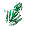
|
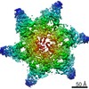
| Symmetry | Helical symmetry: (Circular symmetry: 3 / Dyad axis: no / N subunits divisor: 1 / Num. of operations: 18 / Rise per n subunits: 17.1 Å / Rotation per n subunits: 56.4 °) |
| Details | THE SYMMETRY OF THE HELIX IS D3. |
-Components
| #1: Protein | Mass: 33866.660 Da / Num. of mol.: 2 Source method: isolated from a genetically manipulated source Source: (gene. exp.) Shewanella oneidensis (bacteria) / Strain: MR-1 / Gene: SO_4475 / Production host: |
|---|
-Experimental details
-Experiment
| Experiment | Method: ELECTRON MICROSCOPY |
|---|---|
| EM experiment | Aggregation state: 2D ARRAY / 3D reconstruction method: helical reconstruction |
- Sample preparation
| Component | Name: YiiP from Shewanella oneidensis in DOPG lipids / Type: COMPLEX |
|---|---|
| Buffer solution | Name: 20mM TES, 5mM MgCl2, 100mM NaCl, 5mM NaN3 / pH: 7 / Details: 20mM TES, 5mM MgCl2, 100mM NaCl, 5mM NaN3 |
| Specimen | Conc.: 0.5 mg/ml / Embedding applied: NO / Shadowing applied: NO / Staining applied: NO / Vitrification applied: YES |
| Specimen support | Details: holey carbon grid |
| Vitrification | Instrument: GATAN CRYOPLUNGE 3 / Cryogen name: ETHANE / Temp: 100 K Details: Blot for 2-5 seconds before plunging into liquid ethane (Gatan cryoplunger) Method: Blot for 2-5 seconds before plunging |
- Electron microscopy imaging
| Experimental equipment |  Model: Tecnai F20 / Image courtesy: FEI Company |
|---|---|
| Microscopy | Model: FEI TECNAI F20 / Date: Feb 10, 2009 |
| Electron gun | Electron source:  FIELD EMISSION GUN / Accelerating voltage: 200 kV / Illumination mode: FLOOD BEAM FIELD EMISSION GUN / Accelerating voltage: 200 kV / Illumination mode: FLOOD BEAM |
| Electron lens | Mode: BRIGHT FIELD / Nominal magnification: 50000 X / Calibrated magnification: 51190 X / Nominal defocus max: 3000 nm / Nominal defocus min: 1600 nm / Cs: 2.1 mm |
| Specimen holder | Temperature: 100 K |
| Image recording | Electron dose: 10 e/Å2 / Film or detector model: GENERIC FILM / Details: Kodak SO163 film |
| Image scans | Num. digital images: 19 |
| Radiation | Protocol: SINGLE WAVELENGTH / Monochromatic (M) / Laue (L): M / Scattering type: x-ray |
| Radiation wavelength | Relative weight: 1 |
- Processing
| EM software |
| ||||||||||||
|---|---|---|---|---|---|---|---|---|---|---|---|---|---|
| CTF correction | Details: Corrected throughout the reconstruction cycle | ||||||||||||
| Helical symmerty | Angular rotation/subunit: 56.4 ° / Axial rise/subunit: 17.1 Å / Axial symmetry: D3 | ||||||||||||
| 3D reconstruction | Method: IHRSR / Resolution: 13 Å / Actual pixel size: 2.735 Å Details: CRYSTAL CELL PARAMETERS WERE A=57.5, B=34.0, C=100.0, ALPHA=90, BETA=90, GAMMA=85.3. Symmetry type: HELICAL | ||||||||||||
| Atomic model building | Protocol: FLEXIBLE FIT / Space: REAL Details: METHOD--MDFF DETAILS--An initial homology model of YiiP from S. oneidensis was built with MODELLER 9v7 using the X-ray crystal structure of YiiP from E. coli (PDB entry 3H90) as a template. ...Details: METHOD--MDFF DETAILS--An initial homology model of YiiP from S. oneidensis was built with MODELLER 9v7 using the X-ray crystal structure of YiiP from E. coli (PDB entry 3H90) as a template. This model included 9 residues at the N-terminus and 4 residues at the C-terminus that were not present in the X-ray structure. Initial configurations were obtained by manually placing the symmetry axis of the homology model onto the symmetry axis of the electron density map and aligning the protein C-terminal domains to the corresponding densities. In the first step of the MDFF fitting, harmonic potentials were applied to (a) the four-helix bundle formed by helices M1, M2, M4, and M5 (residues 12-32, 42-65, 119-142, and 147-165, respectively), (b) the M3 and M6 helices at the dimeric interface (residues 78-108 and 179-211, respectively), and (c) the C-terminal intracellular domain (residues 212-297). | ||||||||||||
| Atomic model building | PDB-ID: 3H90 Accession code: 3H90 / Source name: PDB / Type: experimental model | ||||||||||||
| Refinement step | Cycle: LAST
|

