 Movie
Movie Controller

[English] 日本語
 Yorodumi
- PDB-3iy9: Leishmania Tarentolae Mitochondrial Large Ribosomal Subunit Model -
+ Open data

- Basic information
| Entry | Database: PDB / ID: 3iy9 | ||||||
|---|---|---|---|---|---|---|---|
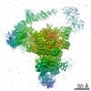
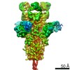
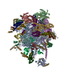
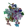
| Title | Leishmania Tarentolae Mitochondrial Large Ribosomal Subunit Model | ||||||
 Components Components |
| ||||||
 Keywords Keywords | RIBOSOME / Leishmania tarentolae / Mitochondrial ribosome / CryoEM / Minimal RNA. / Mitochondrion / Ribonucleoprotein / Ribosomal protein / Transit peptide / Methylation / RNA-binding / rRNA-binding | ||||||
| Function / homology |  Function and homology information Function and homology informationMitochondrial translation elongation / Mitochondrial ribosome-associated quality control / Mitochondrial translation elongation / Mitochondrial translation initiation / Mitochondrial ribosome-associated quality control / Mitochondrial translation termination / mitochondrial large ribosomal subunit / Mitochondrial translation termination / mitochondrial ribosome / mitochondrial translation ...Mitochondrial translation elongation / Mitochondrial ribosome-associated quality control / Mitochondrial translation elongation / Mitochondrial translation initiation / Mitochondrial ribosome-associated quality control / Mitochondrial translation termination / mitochondrial large ribosomal subunit / Mitochondrial translation termination / mitochondrial ribosome / mitochondrial translation / mRNA regulatory element binding translation repressor activity / mRNA 5'-UTR binding / ribosomal large subunit assembly / large ribosomal subunit rRNA binding / cytosolic large ribosomal subunit / cytoplasmic translation / mitochondrial inner membrane / negative regulation of translation / rRNA binding / structural constituent of ribosome / ribosome / translation / protein domain specific binding / mRNA binding / mitochondrion / RNA binding / zinc ion binding / nucleoplasm / cytoplasm / cytosol Similarity search - Function | ||||||
| Biological species |  Leishmania Tarentolae (eukaryote) Leishmania Tarentolae (eukaryote) Homo sapiens (human) Homo sapiens (human) | ||||||
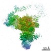
| Method | ELECTRON MICROSCOPY / single particle reconstruction / cryo EM / Resolution: 14.1 Å | ||||||
 Authors Authors | Sharma, M.R. / Booth, T.M. / Simpson, L. / Maslov, D.A. / Agrawal, R.K. | ||||||
 Citation Citation | Journal: Proc Natl Acad Sci U S A / Year: 2009 Title: Structure of a mitochondrial ribosome with minimal RNA. Authors: Manjuli R Sharma / Timothy M Booth / Larry Simpson / Dmitri A Maslov / Rajendra K Agrawal /  Abstract: The Leishmania tarentolae mitochondrial ribosome (Lmr) is a minimal ribosomal RNA (rRNA)-containing ribosome. We have obtained a cryo-EM map of the Lmr. The map reveals several features that have not ...The Leishmania tarentolae mitochondrial ribosome (Lmr) is a minimal ribosomal RNA (rRNA)-containing ribosome. We have obtained a cryo-EM map of the Lmr. The map reveals several features that have not been seen in previously-determined structures of eubacterial or eukaryotic (cytoplasmic or organellar) ribosomes to our knowledge. Comparisons of the Lmr map with X-ray crystallographic and cryo-EM maps of the eubacterial ribosomes and a cryo-EM map of the mammalian mitochondrial ribosome show that (i) the overall structure of the Lmr is considerably more porous, (ii) the topology of the intersubunit space is significantly different, with fewer intersubunit bridges, but more tunnels, and (iii) several of the functionally-important rRNA regions, including the alpha-sarcin-ricin loop, have different relative positions within the structure. Furthermore, the major portions of the mRNA channel, the tRNA passage, and the nascent polypeptide exit tunnel contain Lmr-specific proteins, suggesting that the mechanisms for mRNA recruitment, tRNA interaction, and exiting of the nascent polypeptide in Lmr must differ markedly from the mechanisms deduced for ribosomes in other organisms. Our study identifies certain structural features that are characteristic solely of mitochondrial ribosomes and other features that are characteristic of both mitochondrial and chloroplast ribosomes (i.e., organellar ribosomes). #1: Journal: Science / Year: 2005Title: Structures of the bacterial ribosome at 3.5 A resolution. Authors: Barbara S Schuwirth / Maria A Borovinskaya / Cathy W Hau / Wen Zhang / Antón Vila-Sanjurjo / James M Holton / Jamie H Doudna Cate / Abstract: We describe two structures of the intact bacterial ribosome from Escherichia coli determined to a resolution of 3.5 angstroms by x-ray crystallography. These structures provide a detailed view of the ...We describe two structures of the intact bacterial ribosome from Escherichia coli determined to a resolution of 3.5 angstroms by x-ray crystallography. These structures provide a detailed view of the interface between the small and large ribosomal subunits and the conformation of the peptidyl transferase center in the context of the intact ribosome. Differences between the two ribosomes reveal a high degree of flexibility between the head and the rest of the small subunit. Swiveling of the head of the small subunit observed in the present structures, coupled to the ratchet-like motion of the two subunits observed previously, suggests a mechanism for the final movements of messenger RNA (mRNA) and transfer RNAs (tRNAs) during translocation. #2: Journal: J Mol Biol / Year: 2006Title: A structural model for the large subunit of the mammalian mitochondrial ribosome. Authors: Jason A Mears / Manjuli R Sharma / Robin R Gutell / Amanda S McCook / Paul E Richardson / Thomas R Caulfield / Rajendra K Agrawal / Stephen C Harvey / Abstract: Protein translation is essential for all forms of life and is conducted by a macromolecular complex, the ribosome. Evolutionary changes in protein and RNA sequences can affect the 3D organization of ...Protein translation is essential for all forms of life and is conducted by a macromolecular complex, the ribosome. Evolutionary changes in protein and RNA sequences can affect the 3D organization of structural features in ribosomes in different species. The most dramatic changes occur in animal mitochondria, whose genomes have been reduced and altered significantly. The RNA component of the mitochondrial ribosome (mitoribosome) is reduced in size, with a compensatory increase in protein content. Until recently, it was unclear how these changes affect the 3D structure of the mitoribosome. Here, we present a structural model of the large subunit of the mammalian mitoribosome developed by combining molecular modeling techniques with cryo-electron microscopic data at 12.1A resolution. The model contains 93% of the mitochondrial rRNA sequence and 16 mitochondrial ribosomal proteins in the large subunit of the mitoribosome. Despite the smaller mitochondrial rRNA, the spatial positions of RNA domains known to be involved directly in protein synthesis are essentially the same as in bacterial and archaeal ribosomes. However, the dramatic reduction in rRNA content necessitates evolution of unique structural features to maintain connectivity between RNA domains. The smaller rRNA sequence also limits the likelihood of tRNA binding at the E-site of the mitoribosome, and correlates with the reduced size of D-loops and T-loops in some animal mitochondrial tRNAs, suggesting co-evolution of mitochondrial rRNA and tRNA structures. | ||||||
| History |
|
- Structure visualization
| Movie |
Movie viewer |
|---|---|
| Structure viewer | Molecule: MolmilJmol/JSmol |
- Downloads & links
-Download
| PDBx/mmCIF format | 3iy9.cif.gz | 127.9 KB | Display | PDBx/mmCIF format |
|---|---|---|---|---|
| PDB format | pdb3iy9.ent.gz | 67 KB | Display | PDB format |
| PDBx/mmJSON format | 3iy9.json.gz | Tree view | PDBx/mmJSON format | |
| Others |  Other downloads Other downloads |
-Validation report
| Arichive directory | https://data.pdbj.org/pub/pdb/validation_reports/iy/3iy9ftp://data.pdbj.org/pub/pdb/validation_reports/iy/3iy9 | HTTPS FTP |
|---|
-Related structure data
| Related structure data | 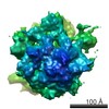 5113MC  3iy8C M: map data used to model this data C: citing same article ( |
|---|---|
| Similar structure data |

-Links
 PDBj
PDBj


- Assembly
| Deposited unit | 
|
|---|---|
| 1 |
|
-Components
-RNA chain , 1 types, 1 molecules A
| #1: RNA chain | Mass: 327983.906 Da / Num. of mol.: 1 / Source method: isolated from a natural source / Source: (natural) Leishmania Tarentolae (eukaryote) |
|---|
-39S ribosomal protein ... , 9 types, 9 molecules BDGISMNOP
| #2: Protein | Mass: 14442.636 Da / Num. of mol.: 1 / Source method: isolated from a natural source / Source: (natural) Homo sapiens (human) / References: UniProt: Q5T653, UniProt: Q2TA12*PLUS |
|---|---|
| #4: Protein | Mass: 19707.887 Da / Num. of mol.: 1 / Source method: isolated from a natural source / Source: (natural) Homo sapiens (human) / References: UniProt: Q9BYD3 |
| #6: Protein | Mass: 15606.364 Da / Num. of mol.: 1 / Source method: isolated from a natural source / Source: (natural) Homo sapiens (human) / References: UniProt: Q9Y3B7, UniProt: Q2YDI0*PLUS |
| #10: Protein | Mass: 13107.179 Da / Num. of mol.: 1 / Source method: isolated from a natural source / Source: (natural) Homo sapiens (human) / References: UniProt: Q9NX20 |
| #11: Protein | Mass: 13682.971 Da / Num. of mol.: 1 / Source method: isolated from a natural source / Source: (natural) Homo sapiens (human) / References: UniProt: Q9NRX2, UniProt: Q3T0L3*PLUS |
| #14: Protein | Mass: 12914.133 Da / Num. of mol.: 1 / Source method: isolated from a natural source / Source: (natural) Homo sapiens (human) / References: UniProt: Q9NWU5, UniProt: Q3SZX5*PLUS |
| #16: Protein | Mass: 11010.680 Da / Num. of mol.: 1 / Source method: isolated from a natural source / Source: (natural) Homo sapiens (human) / References: UniProt: Q96A35, UniProt: Q3SYS0*PLUS |
| #17: Protein | Mass: 7520.631 Da / Num. of mol.: 1 / Source method: isolated from a natural source / Source: (natural) Homo sapiens (human) / References: UniProt: Q9P0M9 |
| #20: Protein | Mass: 6092.276 Da / Num. of mol.: 1 / Source method: isolated from a natural source / Source: (natural) Homo sapiens (human) / References: UniProt: O75394 |
-50S ribosomal protein ... , 10 types, 10 molecules CHJKLQRTXY
| #3: Protein | Mass: 22277.535 Da / Num. of mol.: 1 / Source method: isolated from a natural source / Source: (natural) |
|---|---|
| #5: Protein | Mass: 6580.629 Da / Num. of mol.: 1 / Source method: isolated from a natural source / Source: (natural) |
| #7: Protein | Mass: 15822.360 Da / Num. of mol.: 1 / Source method: isolated from a natural source / Source: (natural) |
| #8: Protein | Mass: 13320.714 Da / Num. of mol.: 1 / Source method: isolated from a natural source / Source: (natural) |
| #9: Protein | Mass: 15008.471 Da / Num. of mol.: 1 / Source method: isolated from a natural source / Source: (natural) |
| #12: Protein | Mass: 13396.828 Da / Num. of mol.: 1 / Source method: isolated from a natural source / Source: (natural) |
| #13: Protein | Mass: 11586.374 Da / Num. of mol.: 1 / Source method: isolated from a natural source / Source: (natural) |
| #15: Protein | Mass: 11093.047 Da / Num. of mol.: 1 / Source method: isolated from a natural source / Source: (natural) |
| #18: Protein | Mass: 7286.464 Da / Num. of mol.: 1 / Source method: isolated from a natural source / Source: (natural) |
| #19: Protein | Mass: 6423.625 Da / Num. of mol.: 1 / Source method: isolated from a natural source / Source: (natural) |
-Experimental details
-Experiment
| Experiment | Method: ELECTRON MICROSCOPY |
|---|---|
| EM experiment | Aggregation state: PARTICLE / 3D reconstruction method: single particle reconstruction |
- Sample preparation
| Component | Name: Leishmania Mitochondrial 50S Ribosome / Type: RIBOSOME / Details: Monomer |
|---|---|
| Molecular weight | Value: 2.1 MDa / Experimental value: YES |
| Buffer solution | Name: 50 mM Tris HCL, pH 7.5, 100mM KCL, 10mM MgCl2, 3mM DTT, 0.1mM EDTA, 0.05% dodecyl maltoside pH: 7.5 Details: 50 mM Tris HCL, pH 7.5, 100mM KCL, 10mM MgCl2, 3mM DTT, 0.1mM EDTA, 0.05% dodecyl maltoside |
| Specimen | Conc.: 0.067 mg/ml / Embedding applied: NO / Shadowing applied: NO / Staining applied: NO / Vitrification applied: YES Details: 50 mM Tris-HCl, pH 7.5, 100 mM KCl, 10 mM MgCl2, 3 mM DTT, 0.1 mM EDTA and 0.05% dodecyl maltoside |
| Specimen support | Details: Thin film carbon |
| Vitrification | Instrument: HOMEMADE PLUNGER / Cryogen name: ETHANE / Humidity: 90 % |
- Electron microscopy imaging
| Experimental equipment |  Model: Tecnai F20 / Image courtesy: FEI Company |
|---|---|
| Microscopy | Model: FEI TECNAI F20 |
| Electron gun | Electron source:  FIELD EMISSION GUN / Accelerating voltage: 200 kV / Illumination mode: FLOOD BEAM FIELD EMISSION GUN / Accelerating voltage: 200 kV / Illumination mode: FLOOD BEAM |
| Electron lens | Mode: BRIGHT FIELD / Nominal magnification: 50000 X / Calibrated magnification: 50760 X / Nominal defocus max: 4500 nm / Nominal defocus min: 1600 nm / Camera length: 0 mm |
| Specimen holder | Specimen holder model: OTHER / Specimen holder type: eucentric / Temperature: 80 K / Tilt angle max: 0 ° / Tilt angle min: 0 ° |
| Image recording | Film or detector model: KODAK SO-163 FILM |
| Radiation | Protocol: SINGLE WAVELENGTH / Monochromatic (M) / Laue (L): M / Scattering type: x-ray |
| Radiation wavelength | Relative weight: 1 |
- Processing
| EM software | Name: SPIDER / Category: 3D reconstruction | ||||||||||||
|---|---|---|---|---|---|---|---|---|---|---|---|---|---|
| CTF correction | Details: Each Micrograph | ||||||||||||
| Symmetry | Point symmetry: C1 (asymmetric) | ||||||||||||
| 3D reconstruction | Method: Projection Matching / Resolution: 14.1 Å / Resolution method: FSC 0.5 CUT-OFF / Num. of particles: 53475 / Symmetry type: POINT | ||||||||||||
| Atomic model building | Space: REAL | ||||||||||||
| Refinement step | Cycle: LAST
|
