Movie
Movie Controller


+ Open data

- Basic information
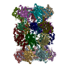
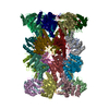
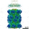
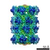
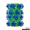
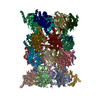
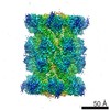
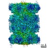
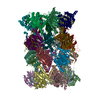
| Entry | Database: PDB / ID: 3c91 | ||||||
|---|---|---|---|---|---|---|---|
| Title | Thermoplasma acidophilum 20S proteasome with an open gate | ||||||
 Components Components |
| ||||||
 Keywords Keywords | HYDROLASE / protein peptide complex / peptide not modeled. / Protease / Proteasome / Threonine protease | ||||||
| Function / homology |  Function and homology information Function and homology informationproteasome endopeptidase complex / proteasome core complex, beta-subunit complex / threonine-type endopeptidase activity / proteasome core complex, alpha-subunit complex / proteasomal protein catabolic process / endopeptidase activity / ubiquitin-dependent protein catabolic process / cytoplasm Similarity search - Function | ||||||
| Biological species |   Thermoplasma acidophilum (acidophilic) Thermoplasma acidophilum (acidophilic) | ||||||
| Method | ELECTRON MICROSCOPY / single particle reconstruction / cryo EM / Resolution: 6.8 Å | ||||||
 Authors Authors | Rabl, J. / Smith, D.M. / Yu, Y. / Chang, S.C. / Goldberg, A.L. / Cheng, Y. | ||||||
 Citation Citation | Journal: Mol Cell / Year: 2008 Title: Mechanism of gate opening in the 20S proteasome by the proteasomal ATPases. Authors: Julius Rabl / David M Smith / Yadong Yu / Shih-Chung Chang / Alfred L Goldberg / Yifan Cheng /  Abstract: Substrates enter the cylindrical 20S proteasome through a gated channel that is regulated by the ATPases in the 19S regulatory particle in eukaryotes or the homologous PAN ATPase complex in archaea. ...Substrates enter the cylindrical 20S proteasome through a gated channel that is regulated by the ATPases in the 19S regulatory particle in eukaryotes or the homologous PAN ATPase complex in archaea. These ATPases contain a conserved C-terminal hydrophobic-tyrosine-X (HbYX) motif that triggers gate opening upon ATP binding. Using cryo-electron microscopy, we identified the sites in the archaeal 20S where PAN's C-terminal residues bind and determined the structures of the gate in its closed and open forms. Peptides containing the HbYX motif bind to 20S in the pockets between neighboring alpha subunits where they interact with conserved residues required for gate opening. This interaction induces a rotation in the alpha subunits and displacement of a reverse-turn loop that stabilizes the open-gate conformation. This mechanism differs from that of PA26/28, which lacks the HbYX motif and does not cause alpha subunit rotation. These findings demonstrated how the ATPases' C termini function to facilitate substrate entry. | ||||||
| History |
|
- Structure visualization
| Movie |
Movie viewer |
|---|---|
| Structure viewer | Molecule: MolmilJmol/JSmol |
- Downloads & links
-Download
| PDBx/mmCIF format | 3c91.cif.gz | 1.1 MB | Display | PDBx/mmCIF format |
|---|---|---|---|---|
| PDB format | pdb3c91.ent.gz | 926.7 KB | Display | PDB format |
| PDBx/mmJSON format | 3c91.json.gz | Tree view | PDBx/mmJSON format | |
| Others |  Other downloads Other downloads |
-Validation report
| Arichive directory | https://data.pdbj.org/pub/pdb/validation_reports/c9/3c91ftp://data.pdbj.org/pub/pdb/validation_reports/c9/3c91 | HTTPS FTP |
|---|
-Related structure data
| Related structure data | 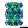 1733MC 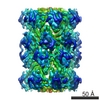 1740C 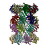 3c92C C: citing same article ( M: map data used to model this data |
|---|---|
| Similar structure data |

-Links
 PDBj
PDBj


- Assembly
| Deposited unit | 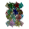
|
|---|---|
| 1 |
|
-Components
| #1: Protein | Mass: 25829.447 Da / Num. of mol.: 14 Source method: isolated from a genetically manipulated source Source: (gene. exp.) Thermoplasma acidophilum (acidophilic) / Production host:  References: UniProt: P25156, proteasome endopeptidase complex #2: Protein | Mass: 22294.848 Da / Num. of mol.: 14 Source method: isolated from a genetically manipulated source Source: (gene. exp.) Thermoplasma acidophilum (acidophilic) / Production host: References: UniProt: P28061, proteasome endopeptidase complex |
|---|
-Experimental details
-Experiment
| Experiment | Method: ELECTRON MICROSCOPY |
|---|---|
| EM experiment | Aggregation state: PARTICLE / 3D reconstruction method: single particle reconstruction |
- Sample preparation
| Component | Name: T. acidophilum 20S proteasome / Type: COMPLEX |
|---|---|
| Buffer solution | Name: 50mM TRIS / pH: 7.5 / Details: 50mM TRIS |
| Specimen | Conc.: 1 mg/ml / Embedding applied: NO / Shadowing applied: NO / Staining applied: NO / Vitrification applied: YES |
| Specimen support | Details: QUANTIFOIL GRID |
| Vitrification | Instrument: FEI VITROBOT MARK I / Cryogen name: NITROGEN / Details: VITROBOT |
- Electron microscopy imaging
| Experimental equipment |  Model: Tecnai F20 / Image courtesy: FEI Company |
|---|---|
| Microscopy | Model: FEI TECNAI F20 / Date: May 1, 2006 / Details: LOW DOSE IMAGING |
| Electron gun | Electron source:  FIELD EMISSION GUN / Accelerating voltage: 200 kV / Illumination mode: FLOOD BEAM FIELD EMISSION GUN / Accelerating voltage: 200 kV / Illumination mode: FLOOD BEAM |
| Electron lens | Mode: BRIGHT FIELD / Nominal magnification: 50000 X / Calibrated magnification: 51159 X / Nominal defocus max: -3500 nm / Nominal defocus min: -1500 nm / Cs: 2 mm |
| Specimen holder | Temperature: 90 K / Tilt angle max: 0 ° / Tilt angle min: 0 ° |
| Image recording | Electron dose: 20 e/Å2 / Film or detector model: KODAK SO-163 FILM |
| Image scans | Num. digital images: 108 |
- Processing
| EM software | Name: FREALIGN / Category: 3D reconstruction | ||||||||||||
|---|---|---|---|---|---|---|---|---|---|---|---|---|---|
| CTF correction | Details: PHASE AND AMPLITUDE | ||||||||||||
| Symmetry | Point symmetry: D7 (2x7 fold dihedral) | ||||||||||||
| 3D reconstruction | Method: FOURIER SPACE / Resolution: 6.8 Å / Num. of particles: 44794 / Nominal pixel size: 1.4 Å / Actual pixel size: 1.37 Å / Magnification calibration: YES Details: GENERATED BY DOCKING THE ATOMIC STRUCTURES OF T. ACIDOPHILUM 20S (1PMA) INTO SINGLE PARTICLE CRYOEM 3D RECONSTRUCTION. PROTEIN PEPTIDE COMPLEX. PEPTIDE WAS NOT MODELED. Symmetry type: POINT | ||||||||||||
| Atomic model building | Protocol: RIGID BODY FIT / Target criteria: CROSS CORRELATION Details: METHOD--USING PROGRAM MAVE IN UPPSALA SOFTWARE FACTOR REFINEMENT PROTOCOL--RIGID BODY | ||||||||||||
| Atomic model building | PDB-ID: 1PMA Accession code: 1PMA / Source name: PDB / Type: experimental model | ||||||||||||
| Refinement | Highest resolution: 6.8 Å | ||||||||||||
| Refinement step | Cycle: LAST / Highest resolution: 6.8 Å
|