+ データを開く
- 基本情報
基本情報
| 登録情報 | データベース: PDB / ID: 3b8k | ||||||
|---|---|---|---|---|---|---|---|
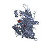
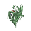
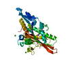
| タイトル | Structure of the Truncated Human Dihydrolipoyl Acetyltransferase (E2) | ||||||
 要素 要素 | Dihydrolipoyllysine-residue acetyltransferase | ||||||
 キーワード キーワード | TRANSFERASE / central beta-sheet surrounded by five alpha-helices | ||||||
| 機能・相同性 |  機能・相同性情報 機能・相同性情報PDH complex synthesizes acetyl-CoA from PYR / dihydrolipoyllysine-residue acetyltransferase / dihydrolipoyllysine-residue acetyltransferase activity / acetyl-CoA biosynthetic process from pyruvate / Regulation of pyruvate dehydrogenase (PDH) complex / Protein lipoylation / pyruvate dehydrogenase complex / Signaling by Retinoic Acid / tricarboxylic acid cycle / glucose metabolic process ...PDH complex synthesizes acetyl-CoA from PYR / dihydrolipoyllysine-residue acetyltransferase / dihydrolipoyllysine-residue acetyltransferase activity / acetyl-CoA biosynthetic process from pyruvate / Regulation of pyruvate dehydrogenase (PDH) complex / Protein lipoylation / pyruvate dehydrogenase complex / Signaling by Retinoic Acid / tricarboxylic acid cycle / glucose metabolic process / mitochondrial matrix / intracellular membrane-bounded organelle / mitochondrion / identical protein binding 類似検索 - 分子機能 | ||||||
| 生物種 |  Homo sapiens (ヒト) Homo sapiens (ヒト) | ||||||
| 手法 | 電子顕微鏡法 / 単粒子再構成法 / クライオ電子顕微鏡法 / 解像度: 8.8 Å | ||||||
 データ登録者 データ登録者 | Yu, X. / Hiromasa, Y. / Tsen, H. / Stoops, J.K. / Roche, T.E. / Zhou, Z.H. | ||||||
 引用 引用 |  ジャーナル: Structure / 年: 2008 ジャーナル: Structure / 年: 2008タイトル: Structures of the human pyruvate dehydrogenase complex cores: a highly conserved catalytic center with flexible N-terminal domains. 著者: Xuekui Yu / Yasuaki Hiromasa / Hua Tsen / James K Stoops / Thomas E Roche / Z Hong Zhou /  要旨: Dihydrolipoyl acetyltransferase (E2) is the central component of pyruvate dehydrogenase complex (PDC), which converts pyruvate to acetyl-CoA. Structural comparison by cryo-electron microscopy (cryo- ...Dihydrolipoyl acetyltransferase (E2) is the central component of pyruvate dehydrogenase complex (PDC), which converts pyruvate to acetyl-CoA. Structural comparison by cryo-electron microscopy (cryo-EM) of the human full-length and truncated E2 (tE2) cores revealed flexible linkers emanating from the edges of trimers of the internal catalytic domains. Using the secondary structure constraints revealed in our 8 A cryo-EM reconstruction and the prokaryotic tE2 atomic structure as a template, we derived a pseudo atomic model of human tE2. The active sites are conserved between prokaryotic tE2 and human tE2. However, marked structural differences are apparent in the hairpin domain and in the N-terminal helix connected to the flexible linker. These permutations away from the catalytic center likely impart structures needed to integrate a second component into the inner core and provide a sturdy base for the linker that holds the pyruvate dehydrogenase for access by the E2-bound regulatory kinase/phosphatase components in humans. | ||||||
| 履歴 |
|
- 構造の表示
構造の表示
| ムービー |
 ムービービューア ムービービューア |
|---|---|
| 構造ビューア | 分子:  Molmil Molmil Jmol/JSmol Jmol/JSmol |
- ダウンロードとリンク
- ダウンロード
| PDBx/mmCIF形式 |  3b8k.cif.gz 3b8k.cif.gz | 52.5 KB | 表示 |  PDBx/mmCIF形式 PDBx/mmCIF形式 |
|---|---|---|---|---|
| PDB形式 |  pdb3b8k.ent.gz pdb3b8k.ent.gz | 36.7 KB | 表示 |  PDB形式 PDB形式 |
| PDBx/mmJSON形式 |  3b8k.json.gz 3b8k.json.gz | ツリー表示 |  PDBx/mmJSON形式 PDBx/mmJSON形式 | |
| その他 |  その他のダウンロード その他のダウンロード |
-検証レポート
| 文書・要旨 |  3b8k_validation.pdf.gz 3b8k_validation.pdf.gz | 896 KB | 表示 |  wwPDB検証レポート wwPDB検証レポート |
|---|---|---|---|---|
| 文書・詳細版 |  3b8k_full_validation.pdf.gz 3b8k_full_validation.pdf.gz | 945.6 KB | 表示 | |
| XML形式データ |  3b8k_validation.xml.gz 3b8k_validation.xml.gz | 23.3 KB | 表示 | |
| CIF形式データ |  3b8k_validation.cif.gz 3b8k_validation.cif.gz | 31.3 KB | 表示 | |
| アーカイブディレクトリ |  https://data.pdbj.org/pub/pdb/validation_reports/b8/3b8k https://data.pdbj.org/pub/pdb/validation_reports/b8/3b8k ftp://data.pdbj.org/pub/pdb/validation_reports/b8/3b8k ftp://data.pdbj.org/pub/pdb/validation_reports/b8/3b8k | HTTPS FTP |
-関連構造データ
- リンク
リンク
- 集合体
集合体
| 登録構造単位 | 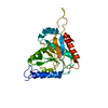
|
|---|---|
| 1 |
|
- 要素
要素
| #1: タンパク質 | 分子量: 25938.164 Da / 分子数: 1 / 断片: C-TERMINAL CATALYTIC DOMAIN / 由来タイプ: 組換発現 詳細: component of pyruvate dehydrogenase complex, mitochondrial 由来: (組換発現)  Homo sapiens (ヒト) / 遺伝子: DLAT, DLTA / 生物種 (発現宿主): Escherichia coli / 発現宿主: Homo sapiens (ヒト) / 遺伝子: DLAT, DLTA / 生物種 (発現宿主): Escherichia coli / 発現宿主:  参照: UniProt: P10515, dihydrolipoyllysine-residue acetyltransferase |
|---|
-実験情報
-実験
| 実験 | 手法: 電子顕微鏡法 |
|---|---|
| EM実験 | 試料の集合状態: PARTICLE / 3次元再構成法: 単粒子再構成法 |
- 試料調製
試料調製
| 構成要素 | 名称: human tE2 / タイプ: COMPLEX 詳細: Dodecahedron. Human tE2 was prepared from scE2, which contains a PreScission site in the third linker region. Treatment of scE2 with the PreScission protease (Amersham Biosciences) removed ...詳細: Dodecahedron. Human tE2 was prepared from scE2, which contains a PreScission site in the third linker region. Treatment of scE2 with the PreScission protease (Amersham Biosciences) removed the N-terminal 319 amino acids. The resulting tE2 was purified by gel filtration with Sephacryl S-300HR. The assembly of the recombinant molecules into fully functional, pentagonal dodecahedral cores was confirmed by analytical ultracentrifugation |
|---|---|
| 緩衝液 | 名称: PBS / pH: 7.2 / 詳細: PBS |
| 試料 | 濃度: 0.2 mg/ml / 包埋: NO / シャドウイング: NO / 染色: NO / 凍結: YES |
| 試料支持 | 詳細: This grid plus sample was kept at -170 deg C during imaging |
| 急速凍結 | 装置: HOMEMADE PLUNGER / 凍結剤: ETHANE / 詳細: flash freezing in liquid ethane |
- 電子顕微鏡撮影
| 顕微鏡 | モデル: JEOL 2010F / 日付: 2003年10月1日 |
|---|---|
| 電子銃 | 電子線源:  FIELD EMISSION GUN / 加速電圧: 200 kV / 照射モード: FLOOD BEAM FIELD EMISSION GUN / 加速電圧: 200 kV / 照射モード: FLOOD BEAM |
| 電子レンズ | モード: BRIGHT FIELD / 倍率(公称値): 69250 X / 倍率(補正後): 69250 X / 最大 デフォーカス(公称値): 2100 nm / 最小 デフォーカス(公称値): 600 nm / Cs: 1 mm |
| 試料ホルダ | 温度: 100 K |
| 撮影 | 電子線照射量: 12 e/Å2 フィルム・検出器のモデル: GATAN ULTRASCAN 4000 (4k x 4k) 詳細: 4kx4k |
- 解析
解析
| EMソフトウェア |
| ||||||||||||
|---|---|---|---|---|---|---|---|---|---|---|---|---|---|
| CTF補正 | 詳細: CTF correction of each image | ||||||||||||
| 対称性 | 点対称性: I (正20面体型対称) | ||||||||||||
| 3次元再構成 | 手法: cross-common lines / 解像度: 8.8 Å / 粒子像の数: 2432 / ピクセルサイズ(公称値): 2.17 Å / ピクセルサイズ(実測値): 2.17 Å 詳細: Orientation determination and 3D reconstruction were performed using the IMIRS software package on multiprocessor MS Windows XP computer workstations. The orientations were first estimated ...詳細: Orientation determination and 3D reconstruction were performed using the IMIRS software package on multiprocessor MS Windows XP computer workstations. The orientations were first estimated from the particle images in the far-from-focus micrographs and refined to about 30- resolution. These orientation parameters were then further refined using the particles in the close-to-focus micrographs. 対称性のタイプ: POINT | ||||||||||||
| 原子モデル構築 | B value: 30 / プロトコル: RIGID BODY FIT / 空間: REAL / Target criteria: best fit using the program CHIMERA / 詳細: REFINEMENT PROTOCOL--rigid body | ||||||||||||
| 原子モデル構築 | PDB-ID: 1EAA Accession code: 1EAA / 詳細: Homology model based on PBD ID 1eaa / Source name: PDB / タイプ: experimental model | ||||||||||||
| 精密化ステップ | サイクル: LAST
|
 ムービー
ムービー コントローラー


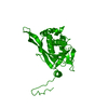
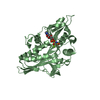

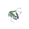


 PDBj
PDBj


