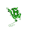
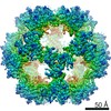
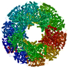
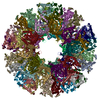
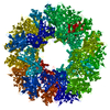
Journal: Structure / Year: 2008 Title: Structures of the human pyruvate dehydrogenase complex cores: a highly conserved catalytic center with flexible N-terminal domains. Authors: Xuekui Yu / Yasuaki Hiromasa / Hua Tsen / James K Stoops / Thomas E Roche / Z Hong Zhou / Abstract: Dihydrolipoyl acetyltransferase (E2) is the central component of pyruvate dehydrogenase complex (PDC), which converts pyruvate to acetyl-CoA. Structural comparison by cryo-electron microscopy (cryo- ...Dihydrolipoyl acetyltransferase (E2) is the central component of pyruvate dehydrogenase complex (PDC), which converts pyruvate to acetyl-CoA. Structural comparison by cryo-electron microscopy (cryo-EM) of the human full-length and truncated E2 (tE2) cores revealed flexible linkers emanating from the edges of trimers of the internal catalytic domains. Using the secondary structure constraints revealed in our 8 A cryo-EM reconstruction and the prokaryotic tE2 atomic structure as a template, we derived a pseudo atomic model of human tE2. The active sites are conserved between prokaryotic tE2 and human tE2. However, marked structural differences are apparent in the hairpin domain and in the N-terminal helix connected to the flexible linker. These permutations away from the catalytic center likely impart structures needed to integrate a second component into the inner core and provide a sturdy base for the linker that holds the pyruvate dehydrogenase for access by the E2-bound regulatory kinase/phosphatase components in humans.
History
Deposition
Nov 1, 2007
-
Header (metadata) release
Nov 1, 2007
-
Map release
Jan 18, 2008
-
Update
May 26, 2011
-
Current status
May 26, 2011
Processing site: PDBe / Status: Released
-
Structure visualization
Movie
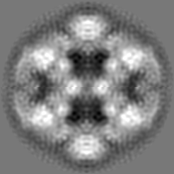
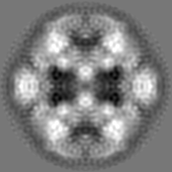
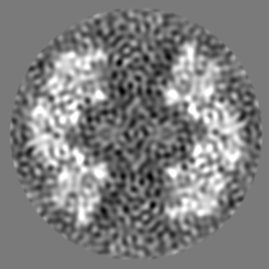
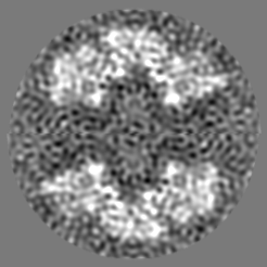
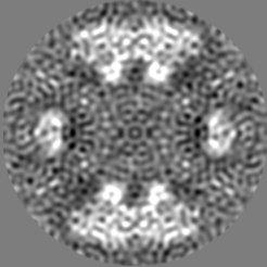
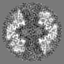
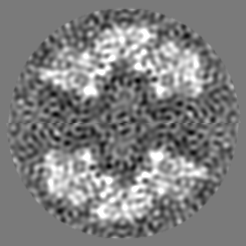
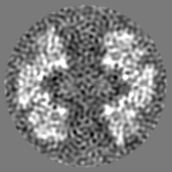
Surface view with section colored by density value
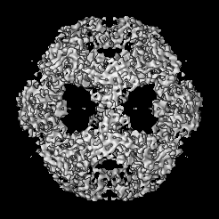
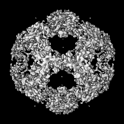
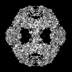
Entire : the truncated human dihydrolipoyl acetyltransferase
Entire
Name: the truncated human dihydrolipoyl acetyltransferase
Components
Sample: the truncated human dihydrolipoyl acetyltransferase
Protein or peptide: truncated human dihydrolipoyl acetyltransferase
-
Supramolecule #1000: the truncated human dihydrolipoyl acetyltransferase
Supramolecule
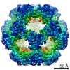
Name: the truncated human dihydrolipoyl acetyltransferase / type: sample / ID: 1000 / Oligomeric state: dodecahedrial assembly of tE2 / Number unique components: 1
Molecular weight
Theoretical: 1.6 MDa
-
Macromolecule #1: truncated human dihydrolipoyl acetyltransferase
Macromolecule
Name: truncated human dihydrolipoyl acetyltransferase / type: protein_or_peptide / ID: 1 / Name.synonym: tE2 Details: Human tE2 was prepared from scE2, which contains a PreScission site in the third linker region. Treatment of scE2 with the PreScission protease (Amersham Biosciences) removed the N-terminal 319 amino acids. Number of copies: 60 / Oligomeric state: Dodecahedron / Recombinant expression: Yes
Source (natural)
Organism: Homo sapiens (human) / synonym: Human
Molecular weight
Experimental: 1.6 MDa / Theoretical: 1.6 MDa
Recombinant expression
Organism: Escherichia coli (E. coli)
-
Experimental details
-
Structure determination
Method
cryo EM
Processing
single particle reconstruction
Aggregation state
particle
-
Sample preparation
Concentration
0.2 mg/mL
Buffer
pH: 7.2 / Details: PBS
Grid
Details: 200 mesh holey carbon grid
Vitrification
Cryogen name: ETHANE / Instrument: HOMEMADE PLUNGER / Details: Vitrification instrument: lab-made plunger / Method: Blot for 1 second before plunging
-
Electron microscopy
Microscope
JEOL 2010F
Temperature
Min: 100 K / Max: 100 K / Average: 100 K
Date
Oct 10, 2003
Image recording
Category: CCD / Film or detector model: GENERIC GATAN / Average electron dose: 12 e/Å2
Electron beam
Acceleration voltage: 200 kV / Electron source: FIELD EMISSION GUN
In the structure databanks used in Yorodumi, some data are registered as the other names, "COVID-19 virus" and "2019-nCoV". Here are the details of the virus and the list of structure data.
Jan 31, 2019. EMDB accession codes are about to change! (news from PDBe EMDB page)
EMDB accession codes are about to change! (news from PDBe EMDB page)
The allocation of 4 digits for EMDB accession codes will soon come to an end. Whilst these codes will remain in use, new EMDB accession codes will include an additional digit and will expand incrementally as the available range of codes is exhausted. The current 4-digit format prefixed with “EMD-” (i.e. EMD-XXXX) will advance to a 5-digit format (i.e. EMD-XXXXX), and so on. It is currently estimated that the 4-digit codes will be depleted around Spring 2019, at which point the 5-digit format will come into force.
The EM Navigator/Yorodumi systems omit the EMD- prefix.
Related info.:Q: What is EMD? / ID/Accession-code notation in Yorodumi/EM Navigator
Yorodumi is a browser for structure data from EMDB, PDB, SASBDB, etc.
This page is also the successor to EM Navigator detail page, and also detail information page/front-end page for Omokage search.
The word "yorodu" (or yorozu) is an old Japanese word meaning "ten thousand". "mi" (miru) is to see.
Related info.:EMDB / PDB / SASBDB / Comparison of 3 databanks / Yorodumi Search / Aug 31, 2016. New EM Navigator & Yorodumi / Yorodumi Papers / Jmol/JSmol / Function and homology information / Changes in new EM Navigator and Yorodumi
 Movie
Movie Controller

 Yorodumi
 Open data
 Basic information
 Map data
 Sample
 Function and homology information
 Homo sapiens (human)
 Authors
 Citation
 Structure visualization
 Downloads & links
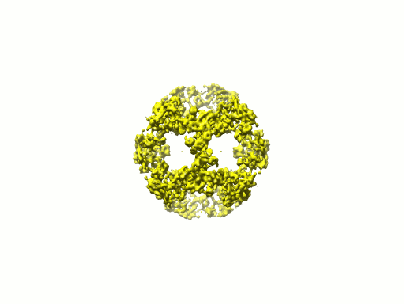 1448.gif
 http://ftp.pdbj.org/pub/emdb/structures/EMD-1448


 Z (Sec.)
 Y (Row.)
 X (Col.)


 Sample components
 Processing
 Electron microscopy
 FIELD EMISSION GUN