Movie
Movie Controller

[English] 日本語
 Yorodumi
- PDB-30gf: Structural characterisation of chromatin remodelling intermediate... -
+ Open data

- Basic information
| Entry | Database: PDB / ID: 30gf | ||||||
|---|---|---|---|---|---|---|---|
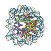
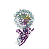
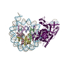
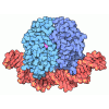
| Title | Structural characterisation of chromatin remodelling intermediates supports linker DNA dependent product inhibition as a mechanism for nucleosome spacing. | ||||||
 Components Components |
| ||||||
 Keywords Keywords | GENE REGULATION / Nucleosome / Remodelling enzyme | ||||||
| Function / homology | DNA / DNA (> 10) / DNA (> 100) Function and homology information Function and homology information | ||||||
| Biological species |   Saccharomyces cerevisiae (brewer's yeast) Saccharomyces cerevisiae (brewer's yeast)synthetic construct (others) | ||||||
| Method | ELECTRON MICROSCOPY / single particle reconstruction / cryo EM / Resolution: 4.8 Å | ||||||
 Authors Authors | Sundaramoorthy, R. / Hughes, A. / Owen-hughes, T.A. | ||||||
| Funding support |  United Kingdom, 1items United Kingdom, 1items
| ||||||
 Citation Citation | Journal: Elife / Year: 2025 Title: Structural characterisation of chromatin remodelling intermediates supports linker DNA-dependent product inhibition as a mechanism for nucleosome spacing. Authors: Amanda L Hughes / Ramasubramanian Sundaramoorthy / Tom Owen-Hughes / Abstract: Previously, we showed that Chd1 chromatin remodelling enzyme associates with nucleosomes oriented towards the longer linker (Sundaramoorthy et al., 2018) (1). Here, we report a series of structures ...Previously, we showed that Chd1 chromatin remodelling enzyme associates with nucleosomes oriented towards the longer linker (Sundaramoorthy et al., 2018) (1). Here, we report a series of structures of Chd1 bound to nucleosomes during ongoing ATP-dependent repositioning. Combining these with biochemical experiments and existing literature, we propose a model in which Chd1 first associates oriented to sample putative entry DNA. In an ATP-dependent reaction, the enzyme then redistributes to the opposite side of the nucleosome, where it subsequently adopts a conformation productive for DNA translocation. Once this active complex extends the nascent exit linker to approximately 15 bp, it is sensed by the Chd1 DNA binding domain, resulting in conversion to a product-inhibited state. These observations provide a mechanistic basis for the action of a molecular ruler element in nucleosome spacing. | ||||||
| History |
|
- Structure visualization
| Structure viewer | Molecule: MolmilJmol/JSmol |
|---|
- Downloads & links
-Download
| PDBx/mmCIF format | 30gf.cif.gz | 476 KB | Display | PDBx/mmCIF format |
|---|---|---|---|---|
| PDB format | pdb30gf.ent.gz | 375.7 KB | Display | PDB format |
| PDBx/mmJSON format | 30gf.json.gz | Tree view | PDBx/mmJSON format | |
| Others |  Other downloads Other downloads |
-Validation report
| Arichive directory | https://data.pdbj.org/pub/pdb/validation_reports/0g/30gfftp://data.pdbj.org/pub/pdb/validation_reports/0g/30gf | HTTPS FTP |
|---|
-Related structure data
| Related structure data |  53597MC  9r5kC  9r5sC  9r5wC M: map data used to model this data C: citing same article ( |
|---|---|
| Similar structure data |


-Links
 PDBj
PDBj
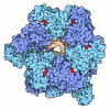



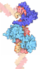







- Assembly
| Deposited unit | 
|
|---|---|
| 1 |
|
-Components
-Protein , 4 types, 8 molecules AEBFCGDH
| #1: Protein | Mass: 15403.062 Da / Num. of mol.: 2 Source method: isolated from a genetically manipulated source Details: structural protein Source: (gene. exp.) Saccharomyces cerevisiae (brewer's yeast)Production host:   Escherichia coli (E. coli) Escherichia coli (E. coli)#2: Protein | Mass: 11394.426 Da / Num. of mol.: 2 Source method: isolated from a genetically manipulated source Details: Structural protein Source: (gene. exp.) Saccharomyces cerevisiae (brewer's yeast)Production host: Escherichia coli (E. coli)#3: Protein | Mass: 13993.295 Da / Num. of mol.: 2 Source method: isolated from a genetically manipulated source Details: structural protein Source: (gene. exp.) Saccharomyces cerevisiae (brewer's yeast)Production host: Escherichia coli (E. coli)#4: Protein | Mass: 13965.265 Da / Num. of mol.: 2 Source method: isolated from a genetically manipulated source Details: structural protein Source: (gene. exp.) Saccharomyces cerevisiae (brewer's yeast)Production host: Escherichia coli (E. coli) |
|---|
-DNA chain , 2 types, 2 molecules IJ
| #5: DNA chain | Mass: 50080.875 Da / Num. of mol.: 1 Source method: isolated from a genetically manipulated source Details: DNA / Source: (gene. exp.) synthetic construct (others) / Production host: synthetic construct (others) |
|---|---|
| #6: DNA chain | Mass: 50557.203 Da / Num. of mol.: 1 Source method: isolated from a genetically manipulated source Details: DNA / Source: (gene. exp.) synthetic construct (others) / Production host: synthetic construct (others) |
-Details
| Has protein modification | N |
|---|
-Experimental details
-Experiment
| Experiment | Method: ELECTRON MICROSCOPY |
|---|---|
| EM experiment | Aggregation state: PARTICLE / 3D reconstruction method: single particle reconstruction |
- Sample preparation
| Component | Name: Nucleosome-Chd1 complex / Type: COMPLEX / Details: Chd1 remodeller bound to Nucleosome / Entity ID: all / Source: RECOMBINANT | |||||||||||||||
|---|---|---|---|---|---|---|---|---|---|---|---|---|---|---|---|---|
| Molecular weight | Value: 0.4 MDa / Experimental value: NO | |||||||||||||||
| Source (natural) | Organism: Saccharomyces cerevisiae (brewer's yeast) | |||||||||||||||
| Source (recombinant) | Organism: Escherichia coli (E. coli) | |||||||||||||||
| Buffer solution | pH: 7.5 / Details: 20mM HEPES, 120mM Nacl pH 7.5 | |||||||||||||||
| Buffer component |
| |||||||||||||||
| Specimen | Conc.: 1.5 mg/ml / Embedding applied: NO / Shadowing applied: NO / Staining applied: NO / Vitrification applied: YES / Details: Purified Nucleosome-Chd1 complex | |||||||||||||||
| Specimen support | Grid material: COPPER/RHODIUM / Grid mesh size: 400 divisions/in. / Grid type: Quantifoil R2/1 | |||||||||||||||
| Vitrification | Instrument: FEI VITROBOT MARK IV / Cryogen name: ETHANE / Humidity: 100 % / Chamber temperature: 277 K Details: Vitrification carried out in climate chamber with 100% humidity |
- Electron microscopy imaging
| Experimental equipment |  Model: Titan Krios / Image courtesy: FEI Company |
|---|---|
| Microscopy | Model: TFS KRIOS |
| Electron gun | Electron source:  FIELD EMISSION GUN / Accelerating voltage: 300 kV / Illumination mode: FLOOD BEAM FIELD EMISSION GUN / Accelerating voltage: 300 kV / Illumination mode: FLOOD BEAM |
| Electron lens | Mode: BRIGHT FIELD / Nominal magnification: 81000 X / Calibrated magnification: 81000 X / Nominal defocus max: 3200 nm / Nominal defocus min: 1800 nm / Calibrated defocus min: 1800 nm / Calibrated defocus max: 3200 nm / Cs: 2.7 mm / C2 aperture diameter: 50 µm / Alignment procedure: COMA FREE |
| Specimen holder | Cryogen: NITROGEN / Specimen holder model: FEI TITAN KRIOS AUTOGRID HOLDER / Temperature (max): 77 K / Temperature (min): 77 K |
| Image recording | Average exposure time: 10 sec. / Electron dose: 46 e/Å2 / Film or detector model: GATAN K3 BIOQUANTUM (6k x 4k) / Num. of grids imaged: 1 / Num. of real images: 2562 |
- Processing
| EM software |
| ||||||||||||||||||||||||||||||||||||||||||||
|---|---|---|---|---|---|---|---|---|---|---|---|---|---|---|---|---|---|---|---|---|---|---|---|---|---|---|---|---|---|---|---|---|---|---|---|---|---|---|---|---|---|---|---|---|---|
| CTF correction | Type: PHASE FLIPPING ONLY | ||||||||||||||||||||||||||||||||||||||||||||
| Particle selection | Num. of particles selected: 2700000 | ||||||||||||||||||||||||||||||||||||||||||||
| 3D reconstruction | Resolution: 4.8 Å / Resolution method: FSC 0.143 CUT-OFF / Num. of particles: 128000 / Algorithm: FOURIER SPACE / Num. of class averages: 1 / Symmetry type: POINT | ||||||||||||||||||||||||||||||||||||||||||||
| Atomic model building | B value: 180 / Protocol: FLEXIBLE FIT / Space: REAL / Target criteria: cross-correlation | ||||||||||||||||||||||||||||||||||||||||||||
| Atomic model building |
| ||||||||||||||||||||||||||||||||||||||||||||
| Refinement | Highest resolution: 4.8 Å Stereochemistry target values: REAL-SPACE (WEIGHTED MAP SUM AT ATOM CENTERS) | ||||||||||||||||||||||||||||||||||||||||||||
| Refine LS restraints |
|