 Movie
Movie Controller

[English] 日本語
 Yorodumi
- PDB-28yj: Molecular basis of ZPD homopolymerization: cryo-EM structure of a... -
+ Open data

- Basic information
| Entry | Database: PDB / ID: 28yj | ||||||||||||||||||||||||||||||
|---|---|---|---|---|---|---|---|---|---|---|---|---|---|---|---|---|---|---|---|---|---|---|---|---|---|---|---|---|---|---|---|
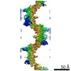
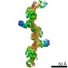
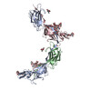
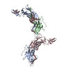
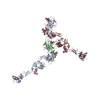
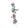
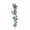
| Title | Molecular basis of ZPD homopolymerization: cryo-EM structure of a native vertebrate egg coat filament | ||||||||||||||||||||||||||||||
 Components Components | Uromodulin | ||||||||||||||||||||||||||||||
 Keywords Keywords | STRUCTURAL PROTEIN / Epidermal growth factor domain / EGF domain / zona pellucida module / zona pellucida domain / ZP module / ZP domain / ZP-N domain / ZP-C domain / interdomain linker / extracellular matrix / glycoprotein / N-glycan / protein filament / protein polymerization / fertilization / egg coat | ||||||||||||||||||||||||||||||
| Function / homology |  Function and homology information Function and homology information: / : / ZP-N domain / Zona pellucida, ZP-C domain / ZP-C domain / Zona pellucida (ZP) domain / ZP domain profile. / Zona pellucida domain / EGF-like domain / Epidermal growth factor-like domain. ...: / : / ZP-N domain / Zona pellucida, ZP-C domain / ZP-C domain / Zona pellucida (ZP) domain / ZP domain profile. / Zona pellucida domain / EGF-like domain / Epidermal growth factor-like domain. / EGF-like domain profile. / EGF-like domain signature 1. / EGF-like domain signature 2. / EGF-like domain Similarity search - Domain/homology | ||||||||||||||||||||||||||||||
| Biological species |  | ||||||||||||||||||||||||||||||
| Method | ELECTRON MICROSCOPY / single particle reconstruction / cryo EM / Resolution: 4.6 Å | ||||||||||||||||||||||||||||||
 Authors Authors | Banjara, S. / Okumura, H. / Jovine, L. | ||||||||||||||||||||||||||||||
| Funding support |  Sweden, 3items Sweden, 3items
| ||||||||||||||||||||||||||||||
 Citation Citation | Journal: Nat Methods / Year: 2026 Title: AlphaFold as a prior: experimental structure determination conditioned on a pretrained neural network. Authors: Alisia Fadini / Minhuan Li / Airlie J McCoy / Suresh Banjara / Hiroki Okumura / Eve Napier / Pietro Fontana / Amir R Khan / Luca Jovine / Thomas C Terwilliger / Randy J Read / Doeke R ...Authors: Alisia Fadini / Minhuan Li / Airlie J McCoy / Suresh Banjara / Hiroki Okumura / Eve Napier / Pietro Fontana / Amir R Khan / Luca Jovine / Thomas C Terwilliger / Randy J Read / Doeke R Hekstra / Mohammed AlQuraishi /     Abstract: Advances in machine learning have transformed structural biology, enabling swift and accurate prediction of protein structure from sequence. However, key challenges persist in modeling side-chain ...Advances in machine learning have transformed structural biology, enabling swift and accurate prediction of protein structure from sequence. However, key challenges persist in modeling side-chain packing, condition-dependent conformational changes and biomolecular interactions, largely because of limited high-quality training data. At the same time, emerging experimental techniques such as cryo-electron microscopy (cryo-EM), cryo-electron tomography (cryo-ET) and high-throughput crystallography are generating vast amounts of structural information but converting these data into mechanistically interpretable atomic models often remains difficult. Here we show that integrating experimental measurements directly into protein structure prediction can overcome these limitations. We introduce ROCKET, an augmentation of AlphaFold2 that refines predicted structures using cryo-EM, cryo-ET and X-ray crystallography data. By optimizing structures in the space of coevolutionary embeddings rather than Cartesian coordinates, ROCKET captures biologically meaningful structural variation that is inaccessible to AlphaFold2 alone and to existing automated modeling approaches, especially when the signal-to-noise ratio is low. ROCKET enables scalable, automated model building without retraining and provides a general framework for integrating experimental observables with biomolecular machine learning. #1: Journal: Biochem J / Year: 2004 Title: A newly identified zona pellucida glycoprotein, ZPD, and dimeric ZP1 of chicken egg envelope are involved in sperm activation on sperm-egg interaction. Authors: Hiroki Okumura / Yoshinori Kohno / Yuki Iwata / Hitoshi Mori / Naohito Aoki / Chihiro Sato / Ken Kitajima / Daita Nadano / Tsukasa Matsuda / Abstract: Fertilization begins with interaction between the sperm and the egg. The surface of the vertebrate oocyte is covered with the egg envelope, which is composed of ZP (zona pellucida) glycoproteins. We ...Fertilization begins with interaction between the sperm and the egg. The surface of the vertebrate oocyte is covered with the egg envelope, which is composed of ZP (zona pellucida) glycoproteins. We have identified two glycoproteins, ZP1/gp97 and ZPC/gp42, as the major components of the chicken egg envelope. In the present study, another 42 kDa protein, designated ZPD, has been found as a new major component of the chicken egg envelope. ZPD was specifically released from the egg envelope by ultrasonication treatment without urea. ZPD cDNA was cloned using a chicken granulosa cell cDNA pool. The deduced amino acid sequence showed that preproprotein of ZPD is composed of 418 amino acid residues with four potential N-glycosylation sites and includes a ZP domain, common in vertebrate ZP glycoproteins, and a transmembrane domain. ZPD belongs phylogenetically to a distinct group from known ZP glycoprotein subfamilies, ZPA, ZPB, and ZPC. In two-dimensional gel electrophoresis ZPD proteins were identified to be several isoforms with different pI values between 5 and 7. ZP1, ZPC and the newly identified ZPD were confirmed to be the major components of chicken egg envelope by MS of proteolytic digests of whole egg envelope. The in vitro incubation of chicken sperm with calcium ionophore A23187 induced sperm activation, resulting in the fragmentation and release of a 41 kDa PNA (peanut agglutinin)-positive glycoprotein and the decrease or loss of sperm PNA-stainability. The incubation with ZPD and dimeric ZP1, but not ZPC and monomeric ZP1, also induced the decrease or loss of sperm PNA-stainability, suggesting the in vitro sperm activation by these ZP components. Collectively, ZPD might bind loosely to egg envelope matrix and play a key role in the sperm activation on avian sperm-egg interaction. #2: Journal: Curr Top Dev Biol / Year: 2018 Title: Egg-Coat and Zona Pellucida Proteins of Chicken as a Typical Species of Aves. Authors: Shunsuke Nishio / Hiroki Okumura / Tsukasa Matsuda / Abstract: Birds are oviparous vertebrates in terrestrial animals. Birds' eggs accumulate mass of egg yolk during the egg development and are accordingly much larger than the eggs of viviparous vertebrates. ...Birds are oviparous vertebrates in terrestrial animals. Birds' eggs accumulate mass of egg yolk during the egg development and are accordingly much larger than the eggs of viviparous vertebrates. Despite such difference in size and contents, the birds' eggs are surrounded with the egg-coat morphologically and compositionally resembling the mammalian egg-coat, zona pellucida. On the other hand, there are some differences in part between the two egg-coats, though relationships of such structural differences to any biological roles specific for the extracellular matrix of birds' eggs are not fully understood. In birds, unlike mammals, ZP proteins constituting the egg-coat are highly conserved and therefore those of chicken are described as a representative of birds. The egg-coat ZP proteins, ZP1, ZP3, and ZPD as the majors, accumulate and form the matrix by self-assembly around the egg rapidly growing in the ovarian follicle, in which ZP1 is from liver and both ZP3 and ZPD are from follicular granulosa cells. Although details of the egg-coat-sperm interaction on fertilization remain to be investigated, the lytic degradation process of egg-coat matrix for the sperm penetration has become to be clarified gradually. ZP1 is the primary target of sperm acrosin, and the limited cleavage in the specific region leading to the loss of intermolecular cross-linkages is crucial for the lysis of egg-coat matrix. Possible roles of the ZP1 with the additional sequence characteristic to birds are discussed from a viewpoint of giving both robustness and elastomeric nature to the egg-coat matrix for the birds' eggs. #3: Journal: EMBO J / Year: 2020Title: Cryo-EM structure of native human uromodulin, a zona pellucida module polymer. Authors: Alena Stsiapanava / Chenrui Xu / Martina Brunati / Sara Zamora-Caballero / Céline Schaeffer / Marcel Bokhove / Ling Han / Hans Hebert / Marta Carroni / Shigeki Yasumasu / Luca Rampoldi / ...Authors: Alena Stsiapanava / Chenrui Xu / Martina Brunati / Sara Zamora-Caballero / Céline Schaeffer / Marcel Bokhove / Ling Han / Hans Hebert / Marta Carroni / Shigeki Yasumasu / Luca Rampoldi / Bin Wu / Luca Jovine /   Abstract: Assembly of extracellular filaments and matrices mediating fundamental biological processes such as morphogenesis, hearing, fertilization, and antibacterial defense is driven by a ubiquitous ...Assembly of extracellular filaments and matrices mediating fundamental biological processes such as morphogenesis, hearing, fertilization, and antibacterial defense is driven by a ubiquitous polymerization module known as zona pellucida (ZP) "domain". Despite the conservation of this element from hydra to humans, no detailed information is available on the filamentous conformation of any ZP module protein. Here, we report a cryo-electron microscopy study of uromodulin (UMOD)/Tamm-Horsfall protein, the most abundant protein in human urine and an archetypal ZP module-containing molecule, in its mature homopolymeric state. UMOD forms a one-start helix with an unprecedented 180-degree twist between subunits enfolded by interdomain linkers that have completely reorganized as a result of propeptide dissociation. Lateral interaction between filaments in the urine generates sheets exposing a checkerboard of binding sites to capture uropathogenic bacteria, and UMOD-based models of heteromeric vertebrate egg coat filaments identify a common sperm-binding region at the interface between subunits. #4: Journal: Cell / Year: 2024Title: ZP2 cleavage blocks polyspermy by modulating the architecture of the egg coat. Authors: Shunsuke Nishio / Chihiro Emori / Benjamin Wiseman / Dirk Fahrenkamp / Elisa Dioguardi / Sara Zamora-Caballero / Marcel Bokhove / Ling Han / Alena Stsiapanava / Blanca Algarra / Yonggang Lu ...Authors: Shunsuke Nishio / Chihiro Emori / Benjamin Wiseman / Dirk Fahrenkamp / Elisa Dioguardi / Sara Zamora-Caballero / Marcel Bokhove / Ling Han / Alena Stsiapanava / Blanca Algarra / Yonggang Lu / Mayo Kodani / Rachel E Bainbridge / Kayla M Komondor / Anne E Carlson / Michael Landreh / Daniele de Sanctis / Shigeki Yasumasu / Masahito Ikawa / Luca Jovine /  Abstract: Following the fertilization of an egg by a single sperm, the egg coat or zona pellucida (ZP) hardens and polyspermy is irreversibly blocked. These events are associated with the cleavage of the N- ...Following the fertilization of an egg by a single sperm, the egg coat or zona pellucida (ZP) hardens and polyspermy is irreversibly blocked. These events are associated with the cleavage of the N-terminal region (NTR) of glycoprotein ZP2, a major subunit of ZP filaments. ZP2 processing is thought to inactivate sperm binding to the ZP, but its molecular consequences and connection with ZP hardening are unknown. Biochemical and structural studies show that cleavage of ZP2 triggers its oligomerization. Moreover, the structure of a native vertebrate egg coat filament, combined with AlphaFold predictions of human ZP polymers, reveals that two protofilaments consisting of type I (ZP3) and type II (ZP1/ZP2/ZP4) components interlock into a left-handed double helix from which the NTRs of type II subunits protrude. Together, these data suggest that oligomerization of cleaved ZP2 NTRs extensively cross-links ZP filaments, rigidifying the egg coat and making it physically impenetrable to sperm. | ||||||||||||||||||||||||||||||
| History |
|
- Structure visualization
| Structure viewer | Molecule: MolmilJmol/JSmol |
|---|
- Downloads & links
-Download
| PDBx/mmCIF format | 28yj.cif.gz | 280.7 KB | Display | PDBx/mmCIF format |
|---|---|---|---|---|
| PDB format | pdb28yj.ent.gz | 229.9 KB | Display | PDB format |
| PDBx/mmJSON format | 28yj.json.gz | Tree view | PDBx/mmJSON format | |
| Others |  Other downloads Other downloads |
-Validation report
| Arichive directory | https://data.pdbj.org/pub/pdb/validation_reports/8y/28yjftp://data.pdbj.org/pub/pdb/validation_reports/8y/28yj | HTTPS FTP |
|---|
-Related structure data
| Related structure data |  56971MC M: map data used to model this data C: citing same article ( |
|---|---|
| Similar structure data |

-Links
 PDBj
PDBj


- Assembly
| Deposited unit | 
|
|---|---|
| 1 |
|
-Components
| #1: Protein | Mass: 36825.234 Da / Num. of mol.: 4 / Source method: isolated from a natural source / Source: (natural) Plasmid details: Zona pellucida (specialized extracellular matrix surrounding the oocyte) Tissue: Oocyte / References: UniProt: Q766V2 #2: Polysaccharide | beta-D-mannopyranose-(1-4)-2-acetamido-2-deoxy-beta-D-glucopyranose-(1-4)-2-acetamido-2-deoxy-beta- ...beta-D-mannopyranose-(1-4)-2-acetamido-2-deoxy-beta-D-glucopyranose-(1-4)-2-acetamido-2-deoxy-beta-D-glucopyranose #3: Polysaccharide | alpha-D-mannopyranose-(1-3)-alpha-D-mannopyranose-(1-6)-[alpha-D-mannopyranose-(1-3)]beta-D- ...alpha-D-mannopyranose-(1-3)-alpha-D-mannopyranose-(1-6)-[alpha-D-mannopyranose-(1-3)]beta-D-mannopyranose-(1-4)-2-acetamido-2-deoxy-beta-D-glucopyranose-(1-4)-2-acetamido-2-deoxy-beta-D-glucopyranose | Source method: isolated from a genetically manipulated source Has ligand of interest | N | Has protein modification | Y | |
|---|
-Experimental details
-Experiment
| Experiment | Method: ELECTRON MICROSCOPY |
|---|---|
| EM experiment | Aggregation state: FILAMENT / 3D reconstruction method: single particle reconstruction |
- Sample preparation
| Component | Name: Native chicken ZPD homopolymeric filament / Type: COMPLEX / Entity ID: #1 / Source: NATURAL |
|---|---|
| Molecular weight | Experimental value: NO |
| Source (natural) | Organism: Cellular location: Zona pellucida (specialized extracellular matrix surrounding the oocyte) Organ: Ovary / Tissue: Oocyte |
| Buffer solution | pH: 7 |
| Buffer component | Conc.: 10 mM / Name: HEPES / Formula: C8H18N2O4S |
| Specimen | Conc.: 0.7 mg/ml / Embedding applied: NO / Shadowing applied: NO / Staining applied: NO / Vitrification applied: YES |
| Vitrification | Cryogen name: NITROGEN |
- Electron microscopy imaging
| Experimental equipment |  Model: Titan Krios / Image courtesy: FEI Company |
|---|---|
| Microscopy | Model: TFS KRIOS |
| Electron gun | Electron source:  FIELD EMISSION GUN / Accelerating voltage: 300 kV / Illumination mode: SPOT SCAN FIELD EMISSION GUN / Accelerating voltage: 300 kV / Illumination mode: SPOT SCAN |
| Electron lens | Mode: BRIGHT FIELD / Nominal magnification: 165000 X / Nominal defocus max: 2800 nm / Nominal defocus min: 700 nm / Cs: 2.7 mm / C2 aperture diameter: 50 µm / Alignment procedure: COMA FREE |
| Specimen holder | Cryogen: NITROGEN / Specimen holder model: FEI TITAN KRIOS AUTOGRID HOLDER |
| Image recording | Average exposure time: 2.75 sec. / Electron dose: 53 e/Å2 / Film or detector model: FEI FALCON IV (4k x 4k) / Num. of real images: 19953 |
| EM imaging optics | Energyfilter name: TFS Selectris / Energyfilter slit width: 10 eV |
- Processing
| EM software |
| ||||||||||||||||||||||||||||||||||||||||||||
|---|---|---|---|---|---|---|---|---|---|---|---|---|---|---|---|---|---|---|---|---|---|---|---|---|---|---|---|---|---|---|---|---|---|---|---|---|---|---|---|---|---|---|---|---|---|
| CTF correction | Type: PHASE FLIPPING AND AMPLITUDE CORRECTION | ||||||||||||||||||||||||||||||||||||||||||||
| Particle selection | Num. of particles selected: 1165059 | ||||||||||||||||||||||||||||||||||||||||||||
| Symmetry | Point symmetry: C1 (asymmetric) | ||||||||||||||||||||||||||||||||||||||||||||
| 3D reconstruction | Resolution: 4.6 Å / Resolution method: FSC 0.143 CUT-OFF / Num. of particles: 498339 / Symmetry type: POINT | ||||||||||||||||||||||||||||||||||||||||||||
| Atomic model building | Protocol: FLEXIBLE FIT / Space: REAL Details: Model building was initiated using a local installation of AlphaFold 3 to predict a minimal filament fragment comprising one full-length subunit (chain A) and two partial subunits (chains B ...Details: Model building was initiated using a local installation of AlphaFold 3 to predict a minimal filament fragment comprising one full-length subunit (chain A) and two partial subunits (chains B and C). The top-ranked prediction was rigid-body fitted into an initial 8.6 A-resolution map (postprocessed with EMReady2) using UCSF Chimera, followed by flexible fitting with Namdinator. Non-resolved terminal regions were trimmed, and well-defined N-glycan densities were manually built in Coot. The model was refined by real-space refinement in Phenix using NCS constraints and increased non-bonded interaction weights, followed by ADP refinement against the unsharpened map. This model served as a starting point for extension with an additional EGF and ZP-N domain from a fourth subunit (chain D). The extended model was docked into the present 4.6 A-resolution map, manually adjusted, and subjected to flexible fitting using the cryo-EM minimizer from cg2all; subsequently, it was refined using Refmac Servalcat task of CCP-EM Doppio, applying global NCS restraints, ProSMART-derived self-restraints, and increased non-bonded interaction weights. Following additional rounds of manual model rebuilding in Coot and real-space refinement in PHENIX (as described above), with positional refinement performed against a LocScale2-postprocessed map and ADP refinement against the unsharpened map, the model was validated using MolProbity and PHENIX. Note that the EGF domain of chain A (and, to a lesser extent, portions of its ZP-N domain near the postprocessed map boundary and the distal regions of the EGF domains in chains C and D) are weakly defined in the density, consistent with their elevated B-factors. These regions were retained in the model to preserve biological completeness, with their conformations constrained by NCS during refinement. | ||||||||||||||||||||||||||||||||||||||||||||
| Atomic model building | Source name: AlphaFold / Type: in silico model | ||||||||||||||||||||||||||||||||||||||||||||
| Refinement | Highest resolution: 4.6 Å Stereochemistry target values: REAL-SPACE (WEIGHTED MAP SUM AT ATOM CENTERS) |
