Movie
Movie Controller


+ Open data

- Basic information
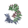
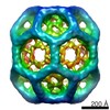
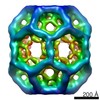
| Entry | Database: PDB / ID: 1xi5 | ||||||
|---|---|---|---|---|---|---|---|
| Title | Clathrin D6 coat with auxilin J-domain | ||||||
 Components Components |
| ||||||
 Keywords Keywords | ENDOCYTOSIS/EXOCYTOSIS / clathrin / alpha-zig-zag / beta-propeller / ENDOCYTOSIS-EXOCYTOSIS COMPLEX | ||||||
| Function / homology |  Function and homology information Function and homology informationregulation of clathrin coat assembly / Retrograde neurotrophin signalling / Recycling pathway of L1 / WNT5A-dependent internalization of FZD4 / WNT5A-dependent internalization of FZD2, FZD5 and ROR2 / LDL clearance / Gap junction degradation / Formation of annular gap junctions / RHOU GTPase cycle / RHOV GTPase cycle ...regulation of clathrin coat assembly / Retrograde neurotrophin signalling / Recycling pathway of L1 / WNT5A-dependent internalization of FZD4 / WNT5A-dependent internalization of FZD2, FZD5 and ROR2 / LDL clearance / Gap junction degradation / Formation of annular gap junctions / RHOU GTPase cycle / RHOV GTPase cycle / clathrin coat of trans-Golgi network vesicle / synaptic vesicle uncoating / Golgi Associated Vesicle Biogenesis / clathrin light chain binding / Lysosome Vesicle Biogenesis / negative regulation of hyaluronan biosynthetic process / clathrin complex / MHC class II antigen presentation / VLDLR internalisation and degradation / synaptic vesicle recycling / clathrin coat of coated pit / clathrin coat disassembly / Cargo recognition for clathrin-mediated endocytosis / clathrin heavy chain binding / clathrin-coated endocytic vesicle / membrane coat / clathrin coat assembly / Hydrolases; Acting on ester bonds; Phosphoric-monoester hydrolases / Clathrin-mediated endocytosis / clathrin-dependent endocytosis / arrestin family protein binding / clathrin-coated vesicle / clathrin binding / intracellular transport / phosphoprotein phosphatase activity / heat shock protein binding / receptor-mediated endocytosis / intracellular protein transport / SH3 domain binding / autophagy / spindle / disordered domain specific binding / melanosome / mitotic cell cycle / presynapse / vesicle / molecular adaptor activity / postsynaptic density / protein domain specific binding / cell division / structural molecule activity / mitochondrion / identical protein binding / cytoplasm Similarity search - Function | ||||||
| Biological species |  | ||||||
| Method | ELECTRON MICROSCOPY / single particle reconstruction / cryo EM / Resolution: 12 Å | ||||||
 Authors Authors | Fotin, A. / Cheng, Y. / Grigorieff, N. / Walz, T. / Harrison, S.C. / Kirchhausen, T. | ||||||
 Citation Citation | Journal: Nature / Year: 2004 Title: Structure of an auxilin-bound clathrin coat and its implications for the mechanism of uncoating. Authors: Alexander Fotin / Yifan Cheng / Nikolaus Grigorieff / Thomas Walz / Stephen C Harrison / Tomas Kirchhausen /  Abstract: Clathrin-coated pits invaginate from specific membrane compartments and pinch off as coated vesicles. These vesicles then uncoat rapidly once released. The Hsc70 molecular chaperone effects the ...Clathrin-coated pits invaginate from specific membrane compartments and pinch off as coated vesicles. These vesicles then uncoat rapidly once released. The Hsc70 molecular chaperone effects the uncoating reaction, and is guided to appropriate locations on clathrin lattices by the J-domain-containing co-chaperone molecule auxilin. This raises the question of how a local event such as ATP hydrolysis by Hsc70 can catalyse a global disassembly. Here, we have used electron cryomicroscopy to determine 12-A-resolution structures of in-vitro-assembled clathrin coats in association with a carboxy-terminal fragment of auxilin that contains both the clathrin-binding region and the J domain. We have located the auxilin fragment by computing differences between these structures and those lacking auxilin (described in an accompanying paper). Auxilin binds within the clathrin lattice near contacts between an inward-projecting C-terminal helical tripod and the crossing of two 'ankle' segments; it also contacts the terminal domain of yet another clathrin 'leg'. It therefore recruits Hsc70 to the neighbourhood of a set of critical interactions. Auxilin binding produces a local change in heavy-chain contacts, creating a detectable global distortion of the clathrin coat. We propose a mechanism by which local destabilization of the lattice promotes general uncoating. #1: Journal: Nature / Year: 2004Title: Molecular model for a complete clathrin lattice from electron cryomicroscopy. Authors: Alexander Fotin / Yifan Cheng / Piotr Sliz / Nikolaus Grigorieff / Stephen C Harrison / Tomas Kirchhausen / Thomas Walz / Abstract: Clathrin-coated vesicles are important vehicles of membrane traffic in cells. We report the structure of a clathrin lattice at subnanometre resolution, obtained from electron cryomicroscopy of coats ...Clathrin-coated vesicles are important vehicles of membrane traffic in cells. We report the structure of a clathrin lattice at subnanometre resolution, obtained from electron cryomicroscopy of coats assembled in vitro. We trace most of the 1,675-residue clathrin heavy chain by fitting known crystal structures of two segments, and homology models of the rest, into the electron microscopy density map. We also define the position of the central helical segment of the light chain. A helical tripod, the carboxy-terminal parts of three heavy chains, projects inward from the vertex of each three-legged clathrin triskelion, linking that vertex to 'ankles' of triskelions centred two vertices away. Analysis of coats with distinct diameters shows an invariant pattern of contacts in the neighbourhood of each vertex, with more variable interactions along the extended parts of the triskelion 'legs'. These invariant local interactions appear to stabilize the lattice, allowing assembly and uncoating to be controlled by events at a few specific sites. | ||||||
| History |
|
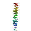
- Structure visualization
| Movie |
Movie viewer |
|---|---|
| Structure viewer | Molecule: MolmilJmol/JSmol |
- Downloads & links
-Download
| PDBx/mmCIF format | 1xi5.cif.gz | 462.3 KB | Display | PDBx/mmCIF format |
|---|---|---|---|---|
| PDB format | pdb1xi5.ent.gz | 285 KB | Display | PDB format |
| PDBx/mmJSON format | 1xi5.json.gz | Tree view | PDBx/mmJSON format | |
| Others |  Other downloads Other downloads |
-Validation report
| Arichive directory | https://data.pdbj.org/pub/pdb/validation_reports/xi/1xi5ftp://data.pdbj.org/pub/pdb/validation_reports/xi/1xi5 | HTTPS FTP |
|---|
-Related structure data
| Related structure data |  5120MC M: map data used to model this data C: citing same article ( |
|---|---|
| Similar structure data |


-Links
 PDBj
PDBj


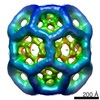
- Assembly
| Deposited unit | 
|
|---|---|
| 1 | x 12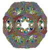
|
| 2 |
|
| 3 | 
|
| Symmetry | Point symmetry: (Hermann–Mauguin notation: 622 / Schoenflies symbol: D6 (2x6 fold dihedral)) |
-Components
| #1: Antibody | Mass: 187145.125 Da / Num. of mol.: 9 / Fragment: residues 1-1630 / Source method: isolated from a natural source / Source: (natural) #2: Protein | Mass: 13383.562 Da / Num. of mol.: 9 / Fragment: residues 797-910 / Source method: isolated from a natural source / Source: (natural) |
|---|
-Experimental details
-Experiment
| Experiment | Method: ELECTRON MICROSCOPY |
|---|---|
| EM experiment | Aggregation state: PARTICLE / 3D reconstruction method: single particle reconstruction |
- Sample preparation
| Component | Name: CLATHRIN D6 COATS WITH BOUND AUXILIN(547-910) / Type: COMPLEX Details: COATS ASSEMBLED WITH LIGHT_CHAIN_FREE CLATHRIN AND AP-2. AUXILIN(547-910) THEN ADDED IN EXCESS |
|---|---|
| Buffer solution | Name: 20MM HEPES / pH: 7 / Details: 20MM HEPES |
| Specimen | Conc.: 1 mg/ml / Embedding applied: NO / Shadowing applied: NO / Staining applied: NO / Vitrification applied: YES |
| Specimen support | Details: HOLEY CARBON |
| Vitrification | Instrument: FEI VITROBOT MARK I / Cryogen name: ETHANE / Details: VITRIFIED |
- Electron microscopy imaging
| Experimental equipment |  Model: Tecnai F20 / Image courtesy: FEI Company |
|---|---|
| Microscopy | Model: FEI TECNAI F20 / Details: LOW DOSE |
| Electron gun | Electron source:  FIELD EMISSION GUN / Accelerating voltage: 200 kV / Illumination mode: FLOOD BEAM FIELD EMISSION GUN / Accelerating voltage: 200 kV / Illumination mode: FLOOD BEAM |
| Electron lens | Mode: BRIGHT FIELD / Nominal magnification: 50000 X / Calibrated magnification: 51160 X / Nominal defocus max: 5000 nm / Nominal defocus min: 2000 nm / Cs: 2 mm |
| Specimen holder | Temperature: 93 K / Tilt angle max: 0 ° / Tilt angle min: 0 ° |
| Image recording | Electron dose: 20 e/Å2 / Film or detector model: KODAK SO-163 FILM |
| Image scans | Num. digital images: 190 |
| Radiation | Protocol: SINGLE WAVELENGTH / Monochromatic (M) / Laue (L): M / Scattering type: x-ray |
| Radiation wavelength | Relative weight: 1 |
- Processing
| EM software |
| ||||||||||||||||||||||||||||
|---|---|---|---|---|---|---|---|---|---|---|---|---|---|---|---|---|---|---|---|---|---|---|---|---|---|---|---|---|---|
| CTF correction | Details: CTFTILT, FREALIGN V.6.07 | ||||||||||||||||||||||||||||
| Symmetry | Point symmetry: D6 (2x6 fold dihedral) | ||||||||||||||||||||||||||||
| 3D reconstruction | Method: FOURIER SPACE RECONSTRUCTION / Resolution: 12 Å / Num. of particles: 900 / Actual pixel size: 2.8 Å Details: The coordinates contain only a CA trace. Please see paper(Nature paper reference) for further details. Symmetry type: POINT | ||||||||||||||||||||||||||||
| Atomic model building | Protocol: OTHER / Space: REAL / Target criteria: DENSITY CORRELATION / Details: METHOD--VISUAL REFINEMENT PROTOCOL--MAVE | ||||||||||||||||||||||||||||
| Atomic model building |
| ||||||||||||||||||||||||||||
| Refinement step | Cycle: LAST
|